- Author Curtis Blomfield blomfield@medicinehelpful.com.
- Public 2023-12-16 20:44.
- Last modified 2025-06-01 06:18.
Our body interacts with the environment through the senses, or analyzers. With their help, a person is not only able to "feel" the outside world, on the basis of these sensations he has special forms of reflection - self-awareness, creativity, the ability to foresee events, etc.
What is an analyzer?
According to IP Pavlov, each analyzer (and even the organ of vision) is nothing but a complex “mechanism”. He is able not only to perceive environmental signals and convert their energy into momentum, but also to produce the highest analysis and synthesis.
The organ of vision, like any other analyzer, consists of 3 integral parts:
- peripheral part, which is responsible for the perception of the energy of external irritation and processing it into a nerve impulse;
- pathways through which the nerve impulse passes directly to the nerve center;
- cortical end of the analyzer (or sensory center) located directly in the brain.
All nerve impulses from analyzers go directly to the central nervous system, where all information is processed. As a result of all these actions, perception arises - the ability to hear, see, touch andetc.
As a sense organ, vision is especially important, because without a bright picture, life becomes boring and uninteresting. It provides 90% of information from the environment.
The eye is an organ of vision that has not yet been fully studied, but still there is an idea of it in anatomy. And this is exactly what will be discussed in the article.

Anatomy and physiology of the organ of vision
Let's take things one at a time.
The organ of vision is the eyeball with the optic nerve and some accessory organs. The eyeball has a spherical shape, usually large in size (its size in an adult is ~ 7.5 cubic cm). It has two poles: back and front. It consists of a nucleus, which is formed by three membranes: fibrous membrane, vascular and retina (or inner membrane). This is the anatomy of the organ of vision. Now about each part in more detail.
Fibrous membrane of the eye
The outer shell of the nucleus consists of the sclera, the posterior region, the dense connective tissue membrane and the cornea, the transparent convex part of the eye, devoid of blood vessels. The cornea is about 1mm thick and about 12mm in diameter.
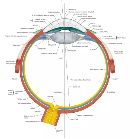
Below is a diagram showing the organ of vision in section. There you can see in more detail where this or that part of the eyeball is located.
Choroid
The second name of this shell of the nucleus is the choroid. It is located directly under the sclera, is saturated with blood vessels and consists of 3 parts: the choroid itself, as well as the iris andciliary body of the eye.
The vascular membrane is a dense network of arteries and veins intertwined. Between them is fibrous loose connective tissue, which is rich in large pigment cells.
In front, the choroid smoothly passes into a thickened ciliary body of an annular shape. Its direct purpose is the accommodation of the eye. The ciliary body supports, fixes and stretches the lens. Consists of two parts: inner (ciliary crown) and outer (ciliary circle).
From the ciliary circle to the lens, about 70 ciliary processes, approximately 2 mm long, depart. The fibers of the zinn ligament (ciliary girdle) are attached to the processes, going to the lens of the eye.
The ciliary girdle consists almost entirely of the ciliary muscle. When it contracts, the lens straightens and rounds, after which its convexity (and with it the refractive power) increases, and accommodation occurs.
Due to the fact that the ciliary muscle cells atrophy in old age and connective tissue cells appear in their place, accommodation deteriorates and farsightedness develops. At the same time, the organ of vision does not cope well with its functions when a person tries to consider something nearby.
Iris
The iris is a round disk with a hole in the center - the pupil. Located between the lens and the cornea.
There are two muscles in the vascular layer of the iris. The first forms the constrictor (sphincter) of the pupil; the second, on the contrary, dilates the pupil.
Exactly fromThe amount of melanin in the iris depends on the color of the eye. Photos of possible options are attached below.

The less pigment in the iris, the lighter the eye color. The organ of vision performs its functions in the same way, regardless of the color of the iris.

Grey-green eye color also means only a small amount of melanin.

The dark color of the eye, the photo of which is higher, indicates that the level of melanin in the iris is high.
Inner (light sensitive) shell
The retina is completely adjacent to the choroid. It is formed by two sheets: outer (pigmented) and inner (light-sensitive).
Three-neuronal radially oriented circuits are isolated in a ten-layer light-sensitive shell, represented by a photoreceptor outer layer, an associative middle layer and a ganglionic inner layer.
Outside, a layer of epithelial pigment cells is attached to the choroid, which are in close contact with the layer of cones and rods. Both are nothing more than peripheral processes (or axons) of photoreceptor cells (neuron I).
Sticks consist of inner and outer segments. The latter is formed with the help of double membrane discs, which are folds of the plasma membrane. Cones differ in size (they are larger) and the nature of the discs.
In the retina, there are three types of cones and only one type of rods. The number of sticks can reach 70million, or even more, while cones are only 5-7 million.
As already mentioned, there are three types of cones. Each of them perceives a different color: blue, red or yellow.
Sticks are needed to perceive information about the shape of an object and the illumination of the room.
From each of the photoreceptor cells, a thin process departs, which forms a synapse (the place where two neurons contact) with another process of bipolar neurons (neuron II). The latter transmit excitation to already larger ganglion cells (neuron III). The axons (processes) of these cells form the optic nerve.
Crystal
This is a biconvex crystal clear lens with a diameter of 7-10mm. It has no nerves or blood vessels. Under the influence of the ciliary muscle, the lens is able to change its shape. It is these changes in the shape of the lens that are called accommodation of the eye. When set to far vision, the lens flattens, and when set to near vision, it increases.
Together with the vitreous body, the lens forms the refractive medium of the eye.
Vitreous body
They fill all the free space between the retina and the lens. Has a jelly-like transparent structure.
The structure of the organ of vision is similar to the principle of the device of the camera. The pupil acts as a diaphragm, constricting or expanding depending on the light. As a lens - the vitreous body and the lens. Light rays strike the retina, but the image is upside down.
body) a beam of light hits the yellow spot on the retina, which is the best zone of vision. Light waves reach cones and rods only after they have passed through the entire thickness of the retina.
Motor apparatus
Motor system of the eye consists of 4 striated rectus muscles (lower, upper, lateral and medial) and 2 oblique (lower and upper). The rectus muscles are responsible for turning the eyeball in the corresponding direction, and the oblique muscles are responsible for turning around the sagittal axis. The movements of both eyeballs are synchronized only thanks to the muscles.
Eyelids
Skin folds, the purpose of which is to limit the palpebral fissure and close it when closed, protect the eyeball from the front. There are about 75 eyelashes on each eyelid, the purpose of which is to protect the eyeball from foreign objects.
Approximately every 5-10 seconds a person blinks.
Lacrimal apparatus
Consists of the lacrimal glands and the lacrimal duct system. Tears neutralize microorganisms and are able to moisten the conjunctiva. Without tears, the conjunctiva of the eye and the cornea would simply dry up and the person would go blind.
The lacrimal glands produce about one hundred milliliters of tears every day. Interesting fact: women cry more than men because the hormone prolactin (which girls have much more) contributes to the release of tear fluid.
Tear is mostly water, containing about 0.5% albumin, 1.5% sodium chloride, some mucus, and lysozyme, which is bactericidal. It has a slightly alkaline reaction.
The structure of the human eye: diagram
Let's take a closer look at the anatomy of the organ of vision with the help of drawings.

The figure above shows schematically parts of the organ of vision in a horizontal section. Here:
1 - tendon of the medial rectus muscle;
2 - rear camera;
3 - cornea;
4 - pupil;
5 - lens;
6 - front camera;
7 - iris;
8 - conjunctiva;
9 - rectus lateralis tendon;
10 - vitreous body;
11 - sclera;
12 - choroid;
13 - retina;
14 - yellow spot;
15 - optic nerve;
16 - retinal blood vessels.

This figure shows a schematic structure of the retina. The arrow shows the direction of the light beam. The numbers are marked:
1 - sclera;
2 - choroid;
3 - retinal pigment cells;
4 - chopsticks;
5 - cones;
6 - horizontal cells;
7 - bipolar cells;
8 - amacrine cells;
9 - ganglion cells;
10 - optic nerve fibers.

The figure shows the scheme of the optical axis of the eye:
1 - object;
2 - cornea;
3 - pupil;
4 - iris;
5 - lens;
6 - center point;
7 - picture.
Whatfunctions performed by the body?
As already mentioned, human vision transmits almost 90% of the information about the world around us. Without him, the world would be the same type and uninteresting.
The organ of vision is a rather complex and not fully understood analyzer. Even in our time, scientists sometimes have questions about the structure and purpose of this organ.
The main functions of the organ of vision are the perception of light, the forms of the surrounding world, the position of objects in space, etc.
Light is able to cause complex changes in the retina of the eye and thus is an adequate irritant for the organs of vision. Rhodopsin is believed to be the first to perceive irritation.
The highest quality visual perception will be provided that the image of the object falls on the area of the retinal spot, preferably on its central fovea. The farther from the center the projection of the image of the object, the less distinct it is. Such is the physiology of the organ of vision.
Diseases of the organ of vision
Let's look at some of the most common eye diseases.
- Hyperopia. The second name for this disease is hypermetropia. A person with this disease does not see objects that are close. It is usually difficult to read, work with small objects. It usually develops in older people, but it can also appear in younger people. Farsightedness can be completely cured only with the help of an operation.
- Myopia (also called myopia). The disease is characterized by the inability to see objects clearly.far enough away.
- Glaucoma is an increase in intraocular pressure. Occurs due to a violation of the circulation of fluid in the eye. It is treated with medication, but in some cases surgery may be required.
- Cataract is nothing more than a violation of the transparency of the lens of the eye. Only an ophthalmologist can help get rid of this disease. Surgery is required to restore a person's vision.
- Inflammatory diseases. These include conjunctivitis, keratitis, blepharitis and others. Each of them is dangerous in its own way and has different methods of treatment: some can be cured with medicines, and some only with the help of operations.
Disease prevention
First of all, you need to remember that your eyes also need to rest, and excessive loads will not lead to anything good.
Use only quality lighting with a 60W to 100W lamp.
Perform eye exercises more often and at least once a year get an examination by an ophthalmologist.
Remember that eye diseases are quite a serious threat to your quality of life.


