- Author Curtis Blomfield blomfield@medicinehelpful.com.
- Public 2024-01-07 17:35.
- Last modified 2025-01-23 17:01.
The foot is the lower part of the lower limb. One side of it, the one that comes into contact with the floor surface, is called the sole, and the opposite, upper side is called the back. The foot has a movable, flexible and elastic vaulted structure with a bulge upwards. The anatomy and shape make it capable of distributing weights, reducing shock when walking, adapting to unevenness, achieving a smooth gait and springy standing.
It performs a supporting function, carries the entire weight of a person and, together with other parts of the leg, moves the body in space.

Foot bones
It is interesting that a quarter of all the bones of his body are located in the feet of a person. So, in one foot there are twenty-six bones. Sometimes it happens that a newborn has more than a few bones. They are called additional and usually they do not hurt their owner.trouble.
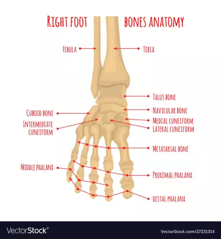
If any bone is damaged, the whole mechanism of the foot will suffer. The anatomy of the bones of the human foot is represented by three sections: tarsus, metatarsus and fingers.
The first section includes seven bones, which are arranged in two rows: the back consists of the calcaneus and the talus, and the anterior consists of the scaphoid, three cuneiform and cuboid.
Each of them has joints that connect them together.
Anatomy of the sole of the foot includes the metatarsus, which includes five short tubular bones. Each of them has a base, a head and a body.
All fingers except the thumb have three phalanges (the thumb has two). All of them are significantly shortened, and on the little finger, the middle phalanx in many people merges with the nail.

Joints of the foot
The anatomy of a joint is represented by two or more interconnected bones. If they get sick, then the strongest pain is felt. Without them, the body would not be able to move, because it is thanks to the joints that the bones can change position relative to each other.
Regarding our topic, the anatomy of the lower leg of the foot is interesting, namely the joint that connects the lower part of the leg to the foot. It has a block shape. When damaged, walking, and even more so running, will cause great pain. Therefore, a person begins to limp, transferring the main weight to the injured leg. This causes the mechanics of both limbs to be broken.
Another in the area under consideration is the subtalar joint, formed from the junction of the posterior calcaneussurfaces with a posterior talar surface. If the foot rotates too much in different directions, it will not work correctly.
But the sphenonavicular joint can compensate for this problem to some extent, especially if it is temporary. However, pathology may eventually occur.
Severe pain, which can be long lasting, occurs in the metatarsophalangeal joints. The greatest pressure falls on the proximal phalanx of the thumb. Therefore, he is the most susceptible to possible pathologies - arthritis, gout and others.
There are other joints in the foot. However, it is the four named that can suffer the most, as they have the maximum impact when walking.
Muscles, joints of the foot
The anatomy of this part is represented by nineteen different muscles, thanks to the interaction of which the leg can move. Overstrain or, on the contrary, underdevelopment will affect them because of the ability to change both the position of the bones and tendons and affect the joints. On the other hand, if something is wrong with the bones, then it will certainly affect the muscles of the foot.
The anatomy of this part of the limb consists of plantar and calf muscles.
Thanks to the first moving toes. Muscles located in different directions help to hold the longitudinal and transverse arches.
The muscles of the lower leg, which are attached by tendons to the bones of the foot, also serve this purpose. These are the anterior and posterior tibial muscles, the long peroneal. From the bones of the lower leg originate those that unbend and bend the fingersfeet. It is important that the muscles of the lower leg and foot are tense. The anatomy of the latter will then be better expressed than with their constantly relaxed state, since otherwise the foot may flatten, which will lead to flat feet.

Tendons and ligaments
Muscles are attached to the bones with the help of tendons, which are their continuation. They are strong, elastic and light. When a muscle is stretched to its limit, force is transferred to the tendon, which can become inflamed if overstretched.
Ligaments are flexible but inelastic tissues. They are located around the joint, supporting it and connecting the bones. When a finger is struck, for example, swelling will be caused by a torn or stretched ligament.
Cartilage
Cartilage covers the ends of bones where joints are located. You can clearly see this white matter at the ends of the chicken leg bone - this is cartilage.
Thanks to him the surfaces of the bones have a smooth appearance. Without cartilage, the body would not be able to move smoothly and the bones would have to knock against each other. In addition, they would feel terrible pain due to their constant inflammation.

Circulatory system
The foot has a dorsal artery and a posterior tibial artery. These are the main arteries that represent the foot. The anatomy of the circulatory system is also represented by smaller arteries, with which they transmit blood and further to all tissues. With insufficient oxygen supply, serious problems arise. These arteries are away from the heartthe strongest. Therefore, circulatory disorders occur primarily in these places. This can be expressed in atherosclerosis and atherosclerosis.
Everyone knows that veins carry blood to the heart. The longest of them runs from the thumb along the entire inner surface of the leg. It is called the great saphenous vein. On the outer side is a small subcutaneous. The anterior and posterior tibials are located deep. Small veins are busy collecting blood from the legs and transferring it to large ones. Small arteries saturate tissues with blood. And capillaries connect arteries and veins.
The image shows the anatomy of the foot. The photo also shows the location of the blood vessels.

Those who have circulatory problems often complain of edema that appears in the afternoon, especially if a lot of time was spent on their feet or after an air flight. Often there is a disease such as varicose veins.
If there is a change in skin color and temperature on the legs, as well as swelling, then these are clear signs that a person has problems with blood circulation. However, the diagnosis should in any case be made by a specialist who should be contacted if the above symptoms are detected.
Nerves
Nerves everywhere transmit sensations to the brain and control the muscles. The foot has the same functions. The anatomy of these formations is represented in it by four types: posterior tibial, deep peroneal, superficial peroneal and sural nerves.
Diseases in this part of the limb can be caused by too much mechanical pressure. For example, tight shoes can compress the nerve, resulting in swelling. This, in turn, will lead to pressure, numbness, pain, or a strange feeling of discomfort.
Functions
After studying the anatomy of the foot, the structure of its individual organs, you can go directly to its functions.
- Due to its mobility, a person easily adapts to the different surface on which he walks. Otherwise, it would have been impossible to do so and he would have simply fallen.
- The body can move in different directions: forward, sideways and backward.
- Most of the load is absorbed by this particular part of the leg. Otherwise, excessive pressure would be created in other parts of her and the body as a whole.

The most common diseases
With a sedentary lifestyle, a disease such as flat feet can develop. It can be transverse and longitudinal.
In the first case, the transverse arch is flattened and the forefoot rests on the heads of all metatarsal bones (in the normal state, it should rest only on the first and fifth). In the second case, the longitudinal arch is flattened, which is why the entire sole is in contact with the surface. With this disease, the legs get tired very quickly and pain is felt in the foot.
Another common disease is osteoarthritis of the ankle joint. There is pain, swelling andcrunch in the specified area. The development of the disease is the damage to the cartilage tissue, which can lead to joint deformity.
No less common is arthrosis of the toes. In this case, there is a violation of blood circulation and metabolic processes in the metatarsophalangeal joints. Symptoms of the disease are pain when moving, crunching, swelling of the fingers, and even the anatomy of the toes (deformity) may be disturbed.
Many people know firsthand what a bump at the base of the thumb is. In official medicine, the disease is called hallux valgus, when the head of the phalangeal bone is displaced. At the same time, the muscles gradually weaken and the thumb begins to lean towards the others, and the foot is deformed.

The anatomy of this part of the lower limb shows its uniqueness and functional importance. Studying the structure of the foot helps to treat it more carefully in order to avoid various diseases.


