- Author Curtis Blomfield blomfield@medicinehelpful.com.
- Public 2024-01-17 01:02.
- Last modified 2025-01-23 17:01.
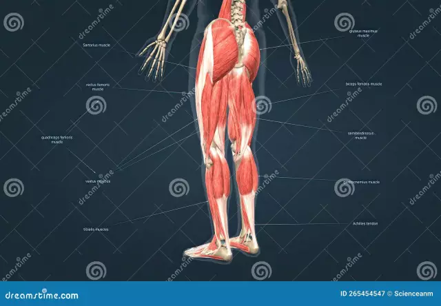
The lower limbs (legs) carry a fairly large load. Their task is to provide movement and support. The muscles of the lower extremities, the anatomy of which will be described in detail in the article, are considered the most powerful of all. Next, consider the muscles of the legs in more detail.

General information
The muscles of the human lower extremities are very well developed. They correct flexion, extension, adduction, abduction of the legs at the knee and hip joint, movement of the fingers and foot. The lower limbs include two muscle groups. The first includes the fibers of the pelvic region. The second group consists of the muscles of the free lower limb. The musculature of the pelvic region begins from the pelvis itself, the lumbar vertebrae and the sacral zone. The fibers are also fixed to the femur. The tasks of the muscles of this part of the leg include keeping the body in a vertical position, extension / flexion of the hip joint and coordination of hip movements. The muscles of the free lower limb include the segments of the thigh, foot and lower leg.
Thigh muscles
The muscles of the human lower extremities in this area are divided into three groups. So,allocate anterior, posterior and medial sections. The first is the flexors, the second is the extensors. The third group includes the muscles that bring the femoral part of the leg. With a significant mass and length, these muscles of the lower extremities of a person can develop great strength. Their activity extends to the knee and hip joints. The thigh muscles perform dynamic and static tasks during walking and standing. Like the segments of the pelvis, these fibers reach their maximum development due to the ability to walk upright.

Muscles of the lower limbs: anatomy. Anterior thigh muscles
It includes the sartorius muscle. The fibers originate from the anterior superior iliac bone. The segment crosses the femoral surface medially, from top to bottom obliquely. The site of attachment is the tuberosity of the tibia and the fascia of the lower leg. At this point, the fibers form a tendon stretch. At the site of attachment, it grows together with similar elements of the semitendinosus and gracilis muscles, forming a fibrous triangular plate - "crow's foot". Underneath is her bag. The functions of this muscle of the lower extremities are to turn the thigh outward, flex it and adduct the lower leg.
Four-headed fibers
They form a strong and large muscle. It has a large mass. The quadriceps muscle includes four segments: intermediate, medial, lateral and direct. From almost all sides, the fibers are adjacent to the femur. In the distal third 4heads form one tendon. It is attached to the tubercle of the tibia, the lateral edges and the apex of the patella.

Straight fibers
They form a muscle starting from the anterior lower iliac bone. Between the fibers and the bone is a synovial bag. The muscle runs down in front of the hip joint. Further, it comes to the surface between the tailor segment and the fibers of the fascia lata. As a result, it occupies a position in front of the wide intermediate muscle. The segment ends with a tendon. It is fixed to the base of the patella. The rectus muscle has a feathery structure.
Lateral segment
This broad thigh muscle is considered the largest of the four. It starts from the intertrochanteric line, gluteal tuberosity, greater trochanter, upper femoral rough line, lateral septum. The fibers are fixed on the tendon of the rectus muscle of the lower limb, the tubercle of the tibia, the upper lateral region of the patella. Part of the tendon bundles continues into the supporting lateral ligament.
Medial segment
This broad muscle has a fairly large beginning. It departs from the lower half of the intertrochanteric, medial lip of the rough line, as well as from the medial femoral septum. The fibers are fixed to the upper end of the base of the patella and the anterior side of the medial condyle on the tibia. The tendon formed by this muscle is involved in the formation of the supporting medial patellar ligament.

Intermediate fibers
They form a broad muscle starting from the upper two-thirds of the lateral and anterior sides of the body of the thigh bone, from the lower part of the lateral lip of the rough line of the thigh and from the lateral intermuscular septum. It is attached to the base of the patella and, together with the tendons of the rectus, lateral and medial wide muscles of the thigh, participates in the formation of the common tendon of the quadriceps femoris.
Shin muscles
She, like other muscles of the girdle of the lower limb, is well developed. This is due to the tasks that she performs. These muscles of the lower limb are associated with dynamics, statics and upright posture. The fibers start extensively on the fasciae, septa, and bones. Their contraction coordinates the movement of the ankle and knee joints. The muscles of the lower limb in this part are divided into lateral, anterior and posterior groups. The latter include the long flexors of the fingers: the big and the rest, popliteal, soleus and gastrocnemius segments. Also in this group is the tibialis posterior muscle. In the anterior section, the long extensors of the fingers are distinguished: the thumb and others. Also present is the tibialis anterior muscle. In the lateral section, long and short peroneal segments are isolated.

Back group
The muscles of this department form deep and superficial layers. The greatest development is noted in the triceps muscle. It lies superficially and forms a characteristicroundness of the leg. The deep layer is formed by a small popliteal and three long muscles: the flexors of the fingers: the thumb and others, as well as the tibialis posterior. They are separated by a plate of the fascia of the leg from the soleus segment.
Lateral group
It is formed by the peroneal muscles of the lower limb: short and long. They run along the lateral side of the leg. These muscles are located between the intermuscular septa (posterior and anterior) under the fascia.
Musculature of the foot
Together with the tendons of the lower leg segments that are fixed to the bones, which belong to the lateral, anterior and posterior groups, there are own (short) fibers in the very lower part of the leg. Their origin and site of attachment is on the skeleton of the foot. Short muscles have complex functional and anatomical and topographic relationships with those tendons of the calf muscles, the fixation points of which are also located on the bones of this part of the leg.

Musculature of the sole of the foot
In this area, medial (in the area of the thumb), lateral (in the area of the little finger) and middle (intermediate) muscle groups are distinguished. On the sole, the first and second sections, in contrast to those on the hand, are represented by a smaller number of fibers. At the same time, the middle muscles on the foot are strengthened. In general, there are 14 short fibers present in the sole. Three segments belong to the medial group, 2 form the lateral one. There are 13 muscles in the middle section: 7 interosseous and 4 worm-like, as well as a square and a short flexor. Significant role in maintaining vaultsis assigned to the muscles not only of the foot itself, but also of the lower leg. Due to this, the tension of the ligamentous apparatus is significantly reduced.
Furrows and channels
Nerves and large vessels of the legs pass through them. In the femoral part they are located between the medial and anterior groups, in the area of the knee joint - in the popliteal fossa, on the sole - between the middle and lateral, as well as between the middle medial sections, on the lower leg - between the muscles of the back surface.

Pelvic muscles of the lower limbs: table
This zone has a practically immovable articulation with the sacral region of the spine. In this regard, the muscles that set it in motion are absent. However, it is these muscles of the human lower extremities that control the activity of the hip joint and spine. The table below summarizes all this information.
| Muscle name | Tasks |
| Iliopumbar | Hip flexion, hip outward rotation |
| Small lumbar | Iliac fascia tension |
| Buttocks Large | Hip leg extension |
| gluteus medius | Hip abduction. With the reduction of internal fibers - rotation inward, rear - outward |
| Buttock minor | Adductionhips. When internal fibers contract, it rotates the thigh inward, the posterior fibers outward |
| Tensor fascia lata | Hip flexion and pronation, fascia lata tension |
| Pear-shaped | Outward hip rotation |
| Internal obturator | |
| Lower and upper twins | |
| External obturator |
Pain in legs
Soreness in the muscles can develop due to various pathologies. These include, in particular:
- Diseases of the spine (sciatica and sciatica, neuritis and neuralgia).
- Pathologies of bones, ligaments and joints (arthritis, arthritis, bursitis, fascia, tendinitis, flat feet, fractures, tumors).
- Direct muscle damage (torn ligaments, myositis, fibromyalgia, cramps, overwork and overexertion).
- Disturbances in metabolic processes and fiber pathology (cellulite, obesity and others).
With paratenonitis and myoenthesitis, pains of a pulling nature appear in the muscles. They arise as a result of inflammatory damage to the fibers and ligaments of the legs. The cause of pathologies is overstrain of muscles against the background of intense loads. Diseases are accompanied by the formation of microtraumas of the muscles and ligaments. Hypothermia, chronic pathologies, general fatigue act as additional risk factors.
In closing
As you know, muscles take an active part in the outflow of blood throughveins. In the process of training the muscles, an increase in the mass of the myocardium is simultaneously carried out. This allows you to carry significant loads. In the process of muscular activity, biologically active compounds, endorphins, are released in the body. They contribute to the adaptation of tissues and organs to a variety of negative influences and provoke a surge of energy and strength. Against the background of physical activity, the organs of the body's defense system are stimulated. In this regard, experts recommend regularly engaging in sports, physical education, doing gymnastic exercises, and taking walks. These activities are of particular importance for the elderly. When playing sports in childhood, the correct posture is formed, the skeleton and muscles develop proportionally.


