- Author Curtis Blomfield blomfield@medicinehelpful.com.
- Public 2023-12-16 20:44.
- Last modified 2025-01-23 17:01.
The basic function of teeth (both in humans and animals) is no secret to anyone. This is the grinding of food (in animals also the capture and retention of prey). The anatomy of the teeth and their shape differ slightly depending on the function performed. There are four types: incisors, canines, premolars and molars. In humans, the first two varieties have a cutting function, and the last ones have a crushing function.

How they work
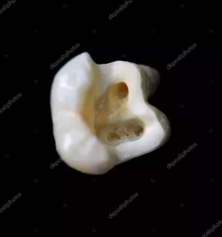
The number of teeth in humans is 32, in animals it is different (depending on the species). They are located in one row on the upper and lower jaws. The structure of the tooth on any of the jaw bones is generally the same, they all consist of similar tissues. Each of them is located in its own bony socket formed by the lower or upper jaw, called the alveolus. Anatomically, the following sections are distinguished in any tooth: crown, neck, root (one or more). The crown is the topmost part accessible for inspection, protruding above the gum. The neck is a small thin section located in the thickness of the soft tissues. Accordingly, the root is the part that goes into the depth of the hole. Its ending is called the tip of the tooth, through which nerves and blood vessels enter the organ. The structure of the tooth (whether canine ormolar) is excellent not only in the external form, but also in the number of roots (from 1 to 3). The shape of the incisors is flat and wide, they have a cutting edge. Canines are characterized by a sharpened crown, premolars and molars - a pronouncedly bumpy chewing surface.

If you describe the internal structure of the tooth, it should be noted that the tissues in it are arranged in layers. The deepest layer - the pulp - is represented by the neurovascular bundle. It is surrounded by dentine, sometimes called the dentary. It is a rather solid substance, however, it can soften when exposed to microorganisms. But explaining the structure of the tooth in its surface layer, it must be said that it is not the same for the crown and root. The first is covered with enamel (an amazingly hard substance, which consists of 97% mineral s alts). The outer layer of the root consists of cement, in which, in addition to lime s alts, there is a large amount of collagen fibers. The latter are woven into the bone tissue, creating the periodontium (and thus attaching the tooth to its socket).
Comparative characteristics

As mentioned above, human teeth have two main functions: for incisors and canines - cutting, for premolars and molars - grinding. In mammals, the last two types perform a similar function. But for incisors and canines it is different. The former are used by predators to “cut off” pieces of food, fangs - to kill and hold the victim. The front cutting teeth of herbivorous living beings are necessary for cutting plants goinginto food. Canines are often absent. If they do exist, they are present only in males. Usually used by them to fight among themselves for a female and leadership in the herd (flock). The microscopic structure of the teeth described above is approximately the same in all species of mammals living organisms.
Now you know what teeth are made of! This information may be useful in some cases!


