- Author Curtis Blomfield blomfield@medicinehelpful.com.
- Public 2023-12-16 20:44.
- Last modified 2025-01-23 17:01.
Various chronic and acute pathologies of the bronchopulmonary system (pneumonia, bronchiectasis, atelectasis, disseminated processes in the lung, cavernous cavities, abscesses, etc.), anemia, and lesions of the nervous system can lead to defects in lung ventilation and the occurrence of respiratory failure., hypertension of the pulmonary circulation, tumors of the mediastinum and lungs, vascular diseases of the heart and lungs, etc.
This article discusses the restrictive type of respiratory failure.

Description of pathology
Restrictive respiratory failure is characterized by a limitation in the ability of lung tissue to collapse and expand, which is observed with pneumothorax, exudative pleurisy, adhesions in the pleural cavity,mobility of the frame of the ribs, kyphoscoliosis, etc. Insufficiency of breathing in such pathologies occurs due to the limitation of the depth of inspiration, which is the maximum possible.
Shapes
Restrictive respiratory failure is caused by defects in alveolar ventilation due to limited lung expansion. There are two forms of ventilatory respiratory failure: pulmonary and extrapulmonary.
Restrictive extrapulmonary ventilation failure develops due to:
- disturbances in the function and structure of the respiratory muscles;
- restrictions (disturbances) of the mobility of the diaphragm and chest;
- increased pressure in the pleural cavity.

Reason
Restrictive respiratory failure must be determined by a doctor. Restrictive pulmonary ventilation insufficiency develops due to a decrease in lung compliance, which is observed during congestive and inflammatory processes. Pulmonary capillaries, overflowing with blood, and interstitial edematous tissue prevent the alveoli from fully expanding, squeezing them. In addition, under these conditions, the extensibility of the interstitial tissue and capillaries decreases.
Symptoms
Restrictive respiratory failure is characterized by a number of symptoms.
- Decrease in lung capacity in general, their residual volume, VC (this indicator reflects the level of pulmonary restriction).
- Defects in the regulatory mechanisms of external respiration. Respiratory disorders also appear due to impaired functioning of the respiratory center, as well as its efferent and afferent connections.
- Manifestation of alveolar restrictive hypoventilation. Clinically significant forms are labored and apneustic breathing, as well as its periodic forms.
- Due to the previous cause and defects in the physico-chemical membrane state, a disorder of the transmembrane ion distribution.
- Violations of neuronal excitability in the respiratory center and, as a result, changes in the depth and frequency of breathing.
- Disorders of external respiratory central regulation. The most common causes are: neoplasms and injuries in the medulla oblongata, compression of the brain (with inflammation or edema, hemorrhages in the medulla or ventricles), intoxication (for example, narcotic drugs, ethanol, endotoxins that are formed during liver failure or uremia), endotoxins, destructive transformations of brain tissue (for example, with syphilis, syringomyelia, multiple sclerosis and encephalitis).
- Defects in the afferent regulation of the activity of the respiratory center, which are manifested by excessive or insufficient afferentation.
- Deficiency of excitatory afferent alveolar restrictive hypoventilation. Reduction of tonic nonspecific activity of neurons located in the reticular formation of the brain stem (acquired or inherited, for example, with an overdose of barbiturates,narcotic analgesics, tranquilizers and other psycho- and neuroactive substances).
- Excessive excitatory afferentation of alveolar restrictive hypoventilation. The signs are as follows: rapid shallow breathing, that is, tachypnea, acidosis, hypercapnia, hypoxia. What is the pathogenesis of restrictive respiratory failure?
- Excessive inhibitory afferentation of alveolar restrictive hypoventilation. The most common causes: increased irritation of the mucous tracts of the respiratory system (when a person inhales irritating substances, for example, ammonia, in acute tracheitis and / or bronchitis while inhaling hot or cold air, severe pain in the airways and / or in the chest (for example, with pleurisy, burns, trauma).
- Defects in the nervous efferent respiratory regulation. Can be observed due to damage at various levels of the effector pathways that regulate the functioning of the respiratory muscles.
- Defects in the cortico-spinal pathways to the muscles of the respiratory system (for example, in syringomyelia, spinal cord ischemia, trauma or tumors), which leads to the loss of conscious (voluntary) control of breathing, as well as the transition to “stabilized”, “machinelike”, “automated” breathing.
- Disorders of the paths leading to the diaphragm from the respiratory center (for example, with spinal cord injury or ischemia, poliomyelitis or multiple sclerosis), which are manifested by a loss of respiratory automatism, as well as a transition tocustom breath.
- Defects in the spinal descending tracts, nerve trunks and motor neurons of the spinal cord to the respiratory muscles (for example, with spinal cord ischemia or trauma, botulism, poliomyelitis, blockade of the conduction of nerves and muscles when using curare and myasthenia gravis, neuritis). Symptoms are as follows: a decrease in the amplitude of respiratory movements and apnea of a periodic nature.


Distinction between restrictive and obstructive respiratory failure
Obstructive respiratory failure, unlike restrictive, is observed when air is difficult to pass through the bronchi and trachea due to bronchospasm, bronchitis (inflammation of the bronchi), penetration of foreign bodies, compression of the trachea and bronchi by a tumor, narrowing (stricture) of the bronchi and trachea etc. At the same time, the functionality of external respiration is violated: a full breath and, in particular, exhalation are difficult, the respiratory rate is limited.
Diagnosis
Restrictive respiratory failure is accompanied by limited air filling of the lungs due to a decrease in the respiratory lung surface, exclusion of part of the lung from breathing, a decrease in the elastic characteristics of the chest and lung, as well as the ability of the lung tissue to stretch (hemodynamic or inflammatory pulmonary edema, extensive pneumonia, pneumosclerosis, pneumoconiosis, etc.). If the restrictive defects are not combined with the bronchial obstruction described above, the resistance of the airways does not increase.

The main consequence of restrictive (restrictive) ventilation disorders, which are detected by classical spirography, is an almost proportional decrease in most of the lung capacities and volumes: FEV1, TO, FEV, VC, ER, ER, etc.
Computer spirography shows that the flow-volume curve is a copy of the correct curve in a reduced form due to a decrease in overall lung volume, which is shifted to the right.
Diagnostic criteria
The most significant diagnostic criteria for restrictive ventilation disorders, which allow quite reliably distinguishing from obstructive defects:
- normal or even elevated Tiffno index (FVC/FEV1);
- virtually proportional decrease in lung capacities and volumes, measured by spirography, and flow indicators, i.e., respectively, a slightly modified or normal shape of the flow-volume loop curve, which is shifted to the right side;
- decrease in EVR (inspiratory reserve volume) is almost proportional to ERV (i.e., expiratory reserve volume).
It should be noted once again that when diagnosing restrictive ventilation disorders in its pure form, one cannot rely only on a decrease in VC. The most reliable diagnostic and differential signs are the absence of transformations in the appearance of the expiratory section of the flow-volume curve and a proportional decrease in ERR andROVD.
How should the patient proceed?
If there are symptoms of restrictive respiratory failure, you need to see a therapist. It may also be necessary to consult specialists from other areas.
Treatment
Restrictive lung disease requires prolonged home ventilation. Her tasks are as follows:
- improving the quality of life;
- extension of human life;
- improving the activity of the respiratory apparatus.
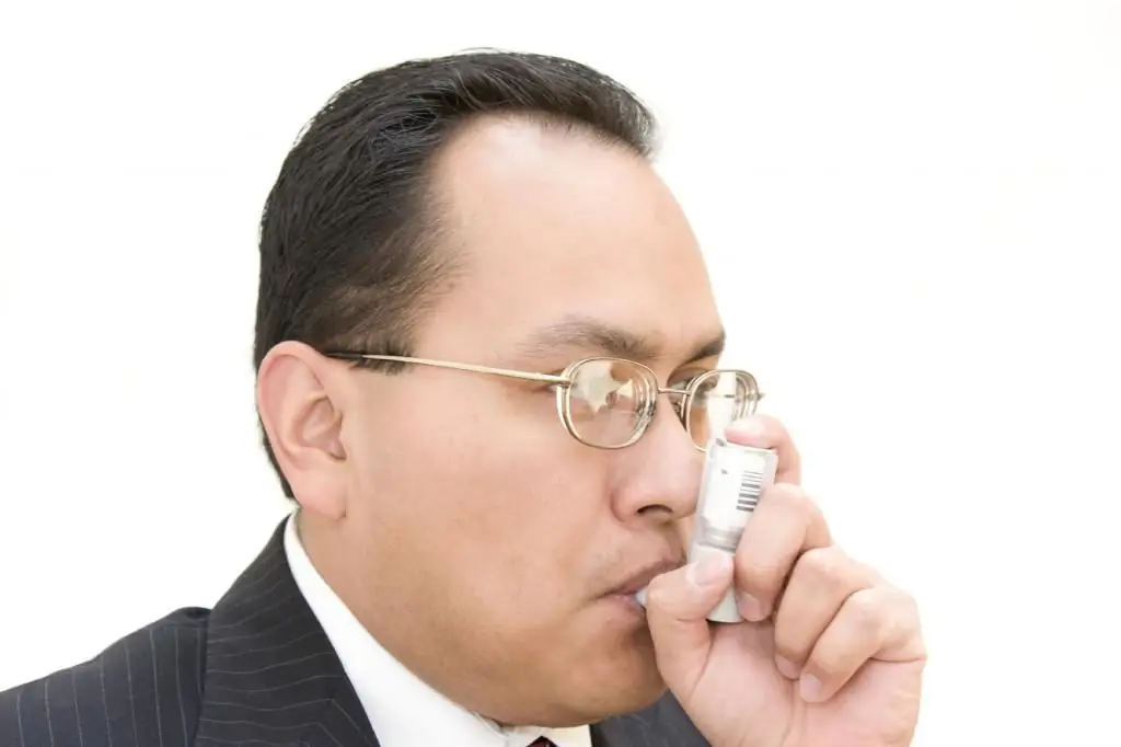
The most common use of long-term home ventilation in patients with restrictive respiratory failure is to use nasal masks and portable respirators (in some cases, a tracheostomy is used), while ventilation is done at night, as well as several hours during the day.
Ventilation parameters are usually selected in stationary conditions, and then the patient is regularly monitored and the equipment is serviced by specialists at home. The most common requirement for long-term home ventilation for patients with chronic respiratory failure is oxygen from liquid oxygen tanks or an oxygen concentrator.
So we looked at restrictive and obstructive types of respiratory failure.


