- Author Curtis Blomfield blomfield@medicinehelpful.com.
- Public 2023-12-16 20:44.
- Last modified 2025-01-23 17:01.
Fractures of the zygomatic bone do not require special procedures to clarify the diagnosis. For experts, stating such damage is not difficult, since it is very easy to recognize.

What characterizes damage?
Damage is characterized by retraction of the bone of the cheekbone, which forms the so-called "step" on the face of the victim. This type of deformity is usually localized in the infraorbital region.
Fractures of the zygomatic bone also make it impossible to fully open the mouth, which is a clear indication of the existing injury. The patient cannot move the lower jaw. At the same time, the fiber of the eye is covered with hemorrhages.
If severe fractures of the zygomatic bone are obtained, then there may be a nosebleed from the nostril located on the affected side.
Usually, for greater certainty, when making a diagnosis, they resort to the use of x-ray equipment, which makes it possible to take a picture showing the damage. Many traumatologists argue that it is extremely difficult to diagnose a fracture of the cheekbone from a picture. But not identifiedfractures can provoke negative consequences leading to pathological changes in the skull area.
What types of zygomatic fractures are there?
As a rule, there are two types of injury: fracture of the zygomatic bone with displacement and fracture of the zygomatic bone without displacement.
Trauma accompanied by displacement is characterized by damage to the maxillary sinuses. It can be closed, open, linear or splinter.
If up to 10 days have passed since the date of the injury, then it is considered fresh, but if more than 10 days or more, then this is an obsolete fracture. If a month has passed since the moment of damage, then the bone is considered to be incorrectly fused or not fused.

Injury symptoms of a displaced fracture
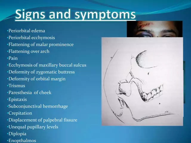
After a fracture of the zygomatic bone, the following symptoms are observed:
- Bleeding, swelling and a wound that masks retraction into the cheekbone area.
- Puffiness of the eyelids that prevents the eyes from closing.
- Frequent bleeding from the nostril on the side of the damaged cheekbone.
- Patient has difficulty opening his mouth. Also, he cannot move his lower jaw in different directions.
- Often there is visual impairment, diplopia associated with the displacement of the eyeball.
- When the zygomatic bone recedes, the patient experiences a sharp pain on palpation.
- Fractures of the zygomatic arch can be combined with fractures of the zygomatic bone. In this case, the formed angle of displacement of bone fragments, as a rule, is directed to the temporal fossa.
Whatis the main task of specialists?
The main task of medical workers during the treatment of an injury is to restore the integrity of the bone. Displaced fractures are eliminated through surgery, since in this case the reduction of bone fragments and their fixation is required. Surgery can take place in and out of the patient's mouth.
Fractures without displacement are treated conservatively with medications and physical therapy.
What complications can occur?
What complications can a zygomatic bone fracture cause? The consequences of a late visit of the victim for medical help may be as follows:
- facial deformity that can become permanent;
- mandibular contracture;
- Chronic upper jaw sinusitis;
- maxillary osteomyelitis.
Mandibular contracture can provoke a displacement of part of the zygomatic bone inward and backward, which contributes to pinching and the development of rough scars in the soft tissues of the coronoid mandibular process.
Chronic maxillary sinusitis and also post-traumatic osteomyelitis provoke bone fragments that are embedded in the sinuses and its gaps.

Treatment of patients with zygomatic injury
How a fracture of the zygomatic bone is repaired. Treatment can be conservative or surgical depending on the extent of the lesion.
When freshinjuries (no more than 10 days from the date of injury) without displaced debris, conservative methods can be used. Rest is usually recommended. Cold is applied to the area of the broken cheekbone. Such measures are carried out within the first two days after the incident.
No pressure on the cheek bone. Mouth opening should be limited as much as possible for two weeks.
Treatment for an old fracture
In case of an old fracture (more than 10 days) with a displacement element, only surgical intervention is indicated. When repositioning bone fragments in the cheekbone, it is contraindicated to open the mouth. With such a lesion, facial deformity, loss of sensitivity to pain in the area of damage to the infraorbital and zygomatic nerves, and double vision are possible.
Fractures of the zygomatic bone and zygomatic arch are eliminated by different methods.

Lumberg method
This is the most commonly used treatment. It is used in the case when damage to the sinus wall is insignificant. A hook with one tooth is used to reduce the bone. The patient assumes a horizontal position. He lies on his back.
The main stages of treatment with the Lamberg method
- The victim's head is on the he althy side.
- A single-pronged hook is inserted through the skin into the area of the displaced zygomatic bone, first in a horizontal direction, and then moves at an acute angle with the tip into the inner surface.
- The fragment is set in the opposite direction to the displacement. Manipulation is carried out until the bone clicks.
Kin's Method
This method is applicable when the zygomatic bone is torn off from the upper jaw, as well as the frontal and temporal bones. First, an incision is made in the mucous membrane in the zone of the transitional fold of the upper jaw behind the alveolar ridge. An elevator is inserted through the wound under the displaced bone by the doctor. By moving up and out, the bone is moved to the correct position.
Vilij's method
It is an improvement on the previous method. It is used to reposition the bone of the cheekbone. The incision is made along the transitional fold in the region of the first and second molars. The Karapetyan elevator is inserted into the bone of the cheekbone or arch, which are repositioned.
Dubov's method
This method is applicable for damage combined with trauma to the walls of the maxillary sinus. How is a fracture of the zygomatic bone repaired in this case? The operation involves making an incision along the upper arch of the mouth from the incisor located in the center to the second molar. The mucous periosteal flap exfoliates, the lateral wall of the upper jaw and sinus are exposed. Fragments of bone are set. Including the bottom of the orbit is affected. An artificial anastomosis is superimposed with the lower course of the nose. The sinus is tightly closed with a swab of gauze soaked in iodoform. Its end is inserted through the nose. The wound located near the mouth is sutured tightly. The tampon is removed after 2 weeks.

Kazanyan-Converse method
This method is similarDubov's method of treatment. But there is some difference. A soft rubber tube is used instead of gauze to hold the bone fragments in the correct position when packing the sinus.
Gillis, Kilner, Stone method
When the cheek bone is broken, a 2 cm incision is made in the temple area. In this case, the doctor steps back from the border of the hairline. A wide Gillis elevator or bent forceps is inserted into the wound. The instrument is advanced into the displaced bone. The tool is supported by a tight gauze swab. Thanks to this manipulation, the fragments can be repositioned.
Duchange Method
With this method, the cheekbone bone is repositioned with forceps specially designed for this purpose with cheeks and sharp teeth. Through the skin with this tool, you can capture the bone of the cheekbone and reposition it. Instead of these tongs, you can use "bullet tongs" or Khodorovich-Barinova tongs.
Malanchuk-Khadarovich treatment method
This method is used for fractures of fresh and old prescription. A hook with one tooth is inserted under the cheek bone or arch and, together with the fragment, is moved outward by means of a lever. The lever rests on the cranial bones.
Osteosynthesis by wire suture or polyamide thread
Fracture of the zygomatic bone, the severity of which is high, is treated with a wire suture. This method is used in the area of the cheekbone and forehead or cheekbone and upper jaw when the fracture gap is exposed in these areas. Small metal plates with small screws are used to fix the fragments of the cheek bone.

Kazanyan's method
This method of treatment is used in the event that the reduction of debris by one manipulation fails, and they cannot be kept in the correct position. The incision is made in the region of the lower eyelid, as a result of which the bone of the cheekbone is exposed in the region of the infraorbital region. A channel is formed in the bone through which a thin stainless wire is passed. The end brought out is bent in the form of a hook or loop. Through this procedure, the zygomatic bone is fixed to the rod, which is mounted in a plaster cap.
Shinbarev's method
The zygomatic bone is fixed with a single-pronged hook to a plaster cast suture bandage. In case of a single fracture of the arc, the hook is inserted strictly along the lower edge at the place where the fragments fall. The skin is sutured. The patient should follow a sparing diet. Cheekbone pressure should be avoided.
Bragin's method
Often, in case of a displaced fracture, it is not possible to fix the fragments in the correct position with a single-pronged hook, since only one fragment of the broken arch is subject to active displacement. In this case, a two-pronged hook is used. There are holes on it through which you can pass under the fragments of the lingature and fix it to the outer tire.
Matas-Berini Method
Using a large curved needle, a thin wire is passed through the tendons in the temporalis muscle above the arch of the cheekbone. The formed loop of wire stretches outward. This way it happensrepositioning of the zygomatic arch.
Matas-Berini Method
This method involves a wire suture. This technique is indicated when other methods do not help. An incision is made along the lower edge of the cheekbone arc, the length of which is 2 cm. The damaged areas are assembled into a single whole. Holes are made at the ends of the fragments with a small burr. With the help of a polyamide thread, the fragments are connected. They are given the correct fixation. The ends of the thread are tied, and the wound is sutured tightly.
In a fracture with many fragments, the bone fragments can be fixed with a plate of fast-hardening plastic. Its width is 1.5 cm, and the length corresponds to the patient's zygomatic arch.
After the fragments are set with a curved needle, a polyamide thread is drawn from the outside under each fragment. The ends of the thread are tied under the plate. A turunda with iodoform is placed between the plate and the skin. This prevents the appearance of bedsores. On day 8-10, the plate is removed.
In the absence of a functional disorder and a long period of time from the day of the fracture (more than 1 year), resection of the coronoid process or osteotomy of the zygomatic bone is used.
Conclusion
Fracture of the zygomatic bone, the photo of which is presented in this article, belongs to the category of severe cases in the field of traumatology.

Untimely treatment of damage can cause a number of undesirable consequences. Therefore, after an injury, it is strongly recommended not topostpone a visit to the doctor. The specialist will prescribe the necessary examination and choose the appropriate treatment method.


