- Author Curtis Blomfield [email protected].
- Public 2023-12-16 20:44.
- Last modified 2025-01-23 17:01.
Bone as an organ is part of the system of organs of movement and support, and at the same time it is distinguished by an absolutely unique shape and structure, a rather characteristic architectonics of nerves and blood vessels. It is built mainly from special bone tissue, which is covered with periosteum on the outside, and contains bone marrow on the inside.
Main Features
Each bone as an organ has a certain size, shape and location in the human body. All this is significantly influenced by the various conditions in which they develop, as well as all kinds of functional loads experienced by the bones throughout the life of the human body.

Any bone is characterized by a certain number of sources of blood supply, the presence of specific places of their location, as well as a rather characteristic architectonics of blood vessels. All these features apply in the same way to the nerves that innervate this bone.
Building
Bone as an organ includes several tissues that are in certain proportions, but, of course, the most important among them is the lamellar bone tissue, the structure of which can be seen on the example of the diaphysis (central section, body) of a long tubular bone.
The main part of it is located betweeninternal and external surrounding plates and is a complex of insertion plates and osteons. The latter is a structural and functional unit of the bone and is examined on specialized histological preparations or thin sections.
Outside, any bone is surrounded by several layers of common or general plates, which are located directly under the periosteum. Through these layers pass specialized perforating channels, which contain blood vessels of the same name. On the border with the medullary cavity, tubular bones also contain an additional layer with internal surrounding plates, pierced by many different channels that expand into cells.
The medullary cavity is entirely lined with the so-called endosteum, which is an extremely thin layer of connective tissue, which includes flattened osteogenic inactive cells.
Osteons
The osteon is represented by concentrically placed bone plates that look like cylinders of different diameters, nested in each other and surrounding the Haversian canal through which various nerves and blood vessels pass. In the vast majority of cases, osteons are placed parallel to the length of the bone, while repeatedly anostomosing with each other.

The total number of osteons is individual for each specific bone. So, for example, the femur as an organ includes them in the amount of 1.8 for every 1 mm², and in this case, the Haversian canal accounts for 0.2-0.3 mm².
Betweenosteons are intermediate or intercalary plates that go in all directions and represent the remaining parts of old osteons that have already collapsed. The structure of the bone as an organ provides for a constant process of destruction and neoformation of osteons.
Bone plates are in the form of cylinders, and ossein fibrils adjoin each other in them tightly and in parallel. Osteocytes are located between concentrically lying plates. The processes of bone cells, gradually spreading through numerous tubules, move towards the processes of neighboring osteocytes and participate in intercellular connections. Thus, they form a spatially oriented lacunar-tubular system, which is directly involved in various metabolic processes.
The composition of the osteon includes more than 20 different concentric bone plates. Human bones pass one or two vessels of the microvasculature through the osteon channel, as well as various unmyelinated nerve fibers and special lymphatic capillaries, which are accompanied by layers of loose connective tissue, which includes various osteogenic elements, such as osteoblasts, perivascular cells and many others.
Osteon channels have a fairly tight connection between themselves, as well as with the medullary cavity and periosteum due to the presence of special awakening channels, which contributes to the overall anastomosis of bone vessels.
Periosteum
The structure of the bone as an organ implies that it is outsidecovered with a special periosteum, which is formed from connective fibrous tissue and has an outer and inner layer. The latter includes cambial progenitor cells.
The main functions of the periosteum include participation in regeneration, as well as providing protective and trophic functions, which is achieved due to the passage of various blood vessels here. Thus, blood and bone interact with each other.
What are the functions of the periosteum
The periosteum almost completely covers the outer part of the bone, and the only exceptions here are the places where the articular cartilage is located, and the ligaments or tendons of the muscles are also fixed. It should be noted that with the help of the periosteum, blood and bone are limited from the surrounding tissues.

In itself, it is an extremely thin, but at the same time strong film, which consists of extremely dense connective tissue, in which the lymphatic and blood vessels and nerves are located. It is worth noting that the latter penetrate into the substance of the bone precisely from the periosteum. Regardless of whether the nasal bone or some other is considered, the periosteum has a fairly large influence on the processes of its development in thickness and nutrition.
The inner osteogenic layer of this coating is the main place where bone tissue is formed, and in itself it is richly innervated, which affects its high sensitivity. If a bone loses its periosteum, it will eventually cease to beviable and completely dead. When carrying out any surgical interventions on the bones, for example, in case of fractures, the periosteum must be preserved without fail in order to ensure their normal further growth and he althy condition.
Other design features
Practically any bones (with the exception of the predominant majority of the cranial, which includes the nasal bone) have articular surfaces that ensure their articulation with others. Instead of a periosteum, such surfaces have specialized articular cartilage, which is fibrous or hyaline in structure.

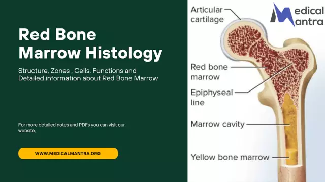
In the vast majority of bones is the bone marrow, which is located between the plates of the spongy substance or is located directly in the medullary cavity, and it can be yellow or red.
In newborns, as well as in fetuses, only red bone marrow is present in the bones, which is hematopoietic and is a homogeneous mass saturated with blood cells, vessels, and also a special reticular tissue. Red bone marrow includes a large number of osteocytes, bone cells. The volume of red bone marrow is approximately 1500 cm³.
In an adult who has already experienced bone growth, the red bone marrow is gradually replaced by yellow, represented mainly by special fat cells, while it is immediately worth noting the fact that only the bone marrow that is located inmedullary cavity.
Osteology
Osteology is concerned with what constitutes the human skeleton, how bones coalesce, and any other processes associated with them. The exact number of described organs in humans cannot be accurately determined because it changes with aging. Few people realize that from childhood to old age, people constantly experience bone damage, tissue death, and many other processes. In general, over 800 different bone elements can develop throughout life, 270 of which are still in the prenatal period.
It is worth noting that the vast majority of them grow together while a person is in childhood and adolescence. In an adult, the skeleton contains only 206 bones, and in addition to permanent bones, in adulthood, non-permanent bones may also appear, the occurrence of which is determined by various individual characteristics and functions of the body.
Skeleton
The bones of the limbs and other parts of the body, together with their joints, form the human skeleton, which is a complex of dense anatomical formations that, in the life of the body, take on mainly exclusively mechanical functions. At the same time, modern science distinguishes a hard skeleton, which appears to be bones, and a soft one, which includes all kinds of ligaments, membranes and special cartilaginous compounds.

Individual bones and joints, as well as the human skeleton inIn general, they can perform a variety of functions in the body. Thus, the bones of the lower extremities and trunk mainly serve as a support for soft tissues, while most bones are levers, since muscles are attached to them, providing locomotor function. Both of these functions make it possible to rightly call the skeleton a completely passive element of the human musculoskeletal system.
The human skeleton is an anti-gravity structure that counteracts the force of gravity. Being under its influence, the human body should be pressed against the ground, but due to the functions that individual bone cells and the skeleton carry in themselves, the body shape does not change.
Bone Functions
The bones of the skull, pelvis and torso provide a protective function against various damage to vital organs, nerve trunks or large vessels:
- the skull is a complete container for the organs of balance, vision, hearing and brain;
- the spinal canal includes the spinal cord;
- the chest provides protection for the lungs, heart, as well as large nerve trunks and blood vessels;
- The pelvic bones protect the bladder, rectum, and various internal genital organs from damage.
The vast majority of bones inside contain red bone marrow, which is a special body of hematopoiesis and the immune system of the human body. It should be noted that the bones protect it from damage, and also createfavorable conditions for the maturation of various formed elements of blood and its trophism.
Among other things, special attention should be paid to the fact that bones are directly involved in mineral metabolism, as they deposit many chemical elements, among which calcium and phosphorus s alts occupy a special place. Thus, if radioactive calcium is introduced into the body, after about 24 hours, more than 50% of this substance will be accumulated in the bones.
Development
Bone is formed by osteoblasts, and there are several types of ossification:
- Endesmal. It is carried out directly in the connective tissue of the integumentary, primary bones. From various points of ossification on the embryo of the connective tissues, the ossification procedure begins to spread in a radiant manner on all sides. The surface layers of the connective tissue remain in the form of a periosteum, from which the bone begins to grow in thickness.
- Perichondral. Occurs on the outer surface of the cartilaginous rudiments with the direct participation of the perichondrium. Thanks to the activity of osteoblasts located under the perichondrium, bone tissue is gradually deposited, replacing cartilage and forming an extremely compact bone substance.
- Periosteal. Occurs due to the periosteum, into which the perichondrium is transformed. The previous and this types of osteogenesis follow each other.
- Endochondral. It is carried out inside the cartilaginous rudiments with the direct participation of the perichondrium, which provides the supplyinside the cartilage processes containing special vessels. This bone-forming tissue gradually destroys the decayed cartilage and forms an ossification point right in the center of the cartilaginous bone model. With further spread of endochondral ossification from the center to the periphery, the formation of spongy bone substance occurs.

How does it happen?
In each person, ossification is functionally determined and begins with the most loaded central parts of the bone. Approximately in the second month of life, primary points begin to appear in the womb, from which the development of the diaphyses, metaphyses and bodies of tubular bones is carried out. In the future, they ossify through endochondral and perichondral osteogenesis, and right before birth or in the first few years after birth, secondary points begin to appear, from which the development of the epiphyses occurs.
In children, as well as people in adolescence and adulthood, additional islands of ossification may appear, from where the development of apophyses begins. Various bones and their individual parts, consisting of a special spongy substance, ossify endochondral over time, while those elements that include spongy and compact substances in their composition ossify peri- and endochondral. The ossification of each individual bone fully reflects its functionally determined processes of phylogenesis.
Height

Throughout growth, there is restructuring and littlebone displacement. New osteons begin to form, and in parallel to this, resorption is also carried out, which is the resorption of all old osteons, which is produced by osteoclasts. Due to their active work, almost completely the entire endochondral bone of the diaphysis eventually resolves, and instead a full-fledged bone marrow cavity is formed. It is also worth noting that the layers of the perichondral bone are also resorbed, and instead of the missing bone tissue, additional layers are deposited from the side of the periosteum. As a result, the bone begins to grow in thickness.
The growth of bones in length is provided by the epiphyseal cartilage, a special layer between the metaphysis and the epiphysis, which persists throughout adolescence and childhood.


