- Author Curtis Blomfield blomfield@medicinehelpful.com.
- Public 2023-12-16 20:44.
- Last modified 2025-01-23 17:01.
Flow cytometry is a cytological research method used for in-depth analysis of cells. Its advantage is that it allows you to study each cell individually. This type of analysis helps to evaluate several parameters in hundreds of cells in a matter of seconds. As a result, cytofluorimetry is considered one of the fastest and most accurate methods of analysis currently available to scientists and clinicians.
Principle
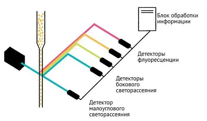
The principle of flow cytometry is based on the measurement of light scattering and luminescence (fluorescence) of cells. The cell suspension is passed as a stream at high speed through the cytometer cell, where it is irradiated with a laser. The so-called hydrodynamic focusing is also performed there. Its mechanism is that the flow from the cell with the studied particles at the outlet flows into the external jet, which has a higher speed. As a result, particles are aligned in an ordered chain.
Pre-cells are labeled with special fluorescent dyes (fluorochromes). Thanks to them, the laser beamexcites a secondary glow. The received light signals are registered by detectors. Subsequently, the information is processed using software algorithms that allow you to count individual cell populations that differ in some criteria.
Research with conventional microscopy often fails to distinguish between different cells because they look the same. Cytofluorometry can provide other data (DNA structure integrity), analyze protein expression, cell survival.
Since excitation of fluorochromes requires light beams with different wavelengths, as well as different types of detectors, modern installations are equipped with several detection channels (from 4 to 30). The number of laser emitters can be from 1 to 7. More complex devices allow multi-parameter studies of several properties of particles at once.
Advantages and disadvantages

The benefits of flow cytometry include:
- high processing speed (registration of up to 30 thousand events in 1 second);
- possibility to study a large number of cells (up to 100 million in a sample);
- Quantitating the intensity of fluorescent light;
- analysis of each cell;
- simultaneous study of heterogeneous processes;
- automatic separation of data by cell populations;
- quality visualization of results.
Another feature of this technology is thatthe analyzed particle can be stained with several fluorescent solutions. Thanks to this, a multi-parameter study occurs.
The disadvantages include the complexity of the technical equipment and the need for special sample preparation.
Cytometers
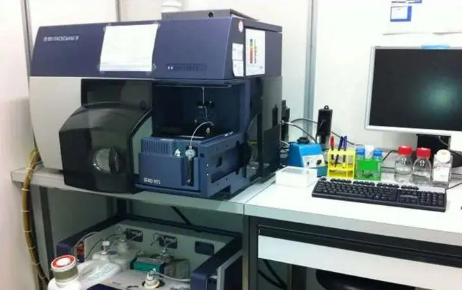
The first devices of this type appeared already in 1968 in Germany, but they became widespread much later. Currently, all devices that work by the method of flow cytometry can be divided into 2 types:
- devices that measure fluorescent radiation (two or more wavelengths), 10° and 90° light scattering (low angle and side scatter detector);
- devices that, in addition to measuring several cellular parameters, automatically sort into groups according to these criteria.
The forward scatter detector is designed to determine the size of the cell, and the side scatter device allows you to obtain information about the presence of intracellular granules, the volume ratio of the cytoplasm and the nucleus.
Classic cytometers, unlike light microscopes, do not allow to obtain an image of a cell. However, in recent years, combined devices have been developed that are able to combine the capabilities of a microscope and a cytofluorimeter. They will be discussed below.
Imaging Cytometers
For instruments used in classical flow cytometry,one feature is characteristic: if rare events are registered in the population of analyzed cells, then there is no way to assess what their essence is. These particles can be either the remains of dead cells or a rare group of them. In conventional devices, such data is excluded from the general flow of events, but it is they that can be of particular value for scientific and clinical analysis.
The new generation of imaging flow cytometers allows you to capture an image of each cell passing in the flow through the detector zone. It is easy to see it by clicking on the corresponding area of the diagram, which is displayed on the computer monitor.
Application areas
Flow cytometry is a universal method that is used in many fields of medicine and science:
- immunology;
- oncology;
- transplantology (transplantation of red bone marrow, stem cells);
- hematology;
- toxicology;
- biochemistry (measurement of acidity inside the cell, the study of other parameters);
- pharmacology (creating new drugs);
- microbiology;
- parasitology and virology;
- oceanology (the study of phytoplankton to assess the state of water bodies and other tasks);
- nanotechnology and microparticle analysis.
Immunology
The human immune system consists of a wide variety of cells. Flow cytometry in immunology makes it possible to evaluate their structure and functions, that is, to carry out morphofunctionalanalysis.
Such research helps to understand the complex nature of immunity. Cell phenotypes change as a result of activation by antigens, the development of pathologies, and other factors. Cytofluorometry can separate subpopulations of immune cells in a complex mixture and evaluate all their changes over time.
Oncology
One of the most important tasks in oncology is the differentiation of cells according to their type. The principle of analysis by flow cytometry in oncohematology is based on the following phenomenon: when a sample is treated with a special fluorescent dye, it binds to cytoplasmic proteins. After division in actively proliferating cells, its content decreases by half. Accordingly, the intensity of cell luminescence decreases twofold.
There are other ways to detect proliferating cells:
- use of DNA-binding dyes (propidium iodide);
- use of labeled uracil;
- registration of an increased level of expression of cyclin proteins, which are involved in the regulation of the cell cycle.


