- Author Curtis Blomfield [email protected].
- Public 2023-12-16 20:44.
- Last modified 2025-01-23 17:01.
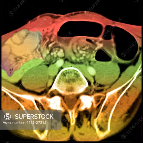
Faced with a complex abbreviation, the patient often asks the doctor what is CT? Deciphering in medicine using professional terminology is presented too complicated. In fact, this is an informative X-ray type diagnostic method with more advanced technology. With it, you can get a three-dimensional image of an organ and a view of a millimeter section of tissue in good quality. This is the simplest explanation of what RCT is.

When it is advisable to use RCT
Before you get acquainted in more detail with the question of what RCT is, you need to understand that in some cases the benefits of its use significantly outweigh the harm. The speed of obtaining information allows you to quickly establish a diagnosis and save lives when there is no time for lengthy laboratory tests.
Through the instrumental method, they reveal:
- Pathologies of the peritoneal organs, hypertrophy of the lymph nodes, inflammatory foci, growths of neoplasms. In a short time, the computer will process the information and determine the volume of the problem, the location.
- Bleeding in the liver, tumors, cysts, dystrophic phenomena, a factor that provoked the formation of jaundice.
- Evaluation of tissue density by CT scan of the brain, which reveals aneurysms, tumors, predisposition and consequences of stroke.
- Diseases of the chest organs (pneumonia, cancer, life processes of Koch's bacillus), the state of blood flow, the heart muscle, it is possible to identify stenosis of the esophagus, destructive changes in the thoracic spine. CT scan of the lungs detects any anomalies in the alveoli, which allows stopping the processes at the initial stages.
- Changes in the spine, hernias, fissures, fractures, foci of infections. Determine diseases of bones, joints, muscle fibers of the limbs.
- Assess the condition of the kidneys and ureters. The device detects stones, neoplasms, anomalies of congenital nature.
- Intestinal diseases, used as a control method to prevent cancer after 50 years.
- Pathological changes in the reproductive organs.
It is worth noting that initially doctors resort to simpler and safer diagnostic methods. If the cause of the manifestation of symptoms can be established by laboratory research methods or other instrumental methods,then they are used. In order to clarify the result, there may be an indication of CT diagnostics.
Doctor may order CT if needed:
- carry out a screening test (chronic diseases, suspected oncology);
- immediately diagnose the cause of seizures and bleeding, see a clear picture of the consequences of injuries;
- additional surveys due to lack of information previously conducted;
- specify the location of the pathological focus before surgery.
The method is quite expensive, the attending physician prescribes it only when there is no way around it.
Advantages of the diagnostic method
Compared to radiography, CT wins. The main advantages of the modern method are:
- 20 times the expansion capacity of X-rays;
- no blurring;
- getting a picture in three dimensions.
The similarity with x-rays is that both are sources of radiation. The average radiation dose is 2-11 m3v (the figure may vary for various reasons).

Preparing the patient for examination
If the doctor prescribes an x-ray computed tomography of the skull, brain, nose, temples, neck, thyroid gland, larynx, sternum, spinal column, shoulder blades, large articular surfaces - there is no need for preparation. But it is definitely worth studying the topic and understanding what it is - RCT research.
CT of the peritoneum without the use of contrast is done on an empty stomach. If the study is carried out using an intravenous injection of contrast, then they first get acquainted with the patient's allergic history, determine the absence of contraindications to the components of the injected substance. CT with an amplifier is performed on an empty stomach. In addition to the daily rate of water consumed, you should drink up to two liters of fluid the day before the procedure.
RCT colonography requires bowel cleansing. If coronary angiography is planned, control over the following indicators cannot be dispensed with:
- heart contraction at rest - 65 bpm;
- no cardiac arrhythmias;
- the ability to hold your breath for 25 seconds
When examining the liver, spleen, pancreas, kidneys, the study is performed on an empty stomach. A day before the procedure, you should refuse raw vegetables, fruits, juices, water with gases, black bread and milk. Enzyme preparations and antioxidants are not accepted.

How the procedure works
Knowing all the pros and cons of the research method, what is CT and how it is possible to obtain an image in several projections, the whole world continues to use the medical instrumental approach to diagnose serious pathologies.
X-rays, penetrating the tissues, are reflected on the sensors with different strengths. The intensity may vary, as the organs have a different structure. The density of the reverse flow forms an image - a tomogram.
Important! The presence of implants, metal objects is not an obstacle to diagnosis, unlike MRI.
X-ray computed tomography is performed using x-rays. The modern research method allows obtaining a two-dimensional three-dimensional image. In the device device, there is a feature that distinguishes CT from conventional x-rays. The source of the rays is a ring-like contour. There is a couch inside. The position of the patient allows you to receive images from various points at convenient angles. The computer collects information, processes it and produces a 3D model of the organ.
Physicians use one of the three existing types of RCT research:
- The rotation of the X-ray tube around the patient and the simultaneous movement of the patient - spiral. Due to the fact that the procedure is carried out quickly, it is possible to obtain a minimum dose of radiation.
- XCT system that allows scanning up to 500 layers. The multilayer technique receives radiation from sensors arranged in several rows. Thus, you can examine the body in the process.
- Multispiral CT of organs is carried out using accelerated technology: the equipment scans quickly and is able to increase resolution. Small vessels are examined with the help of the device. The diagnostic method is used by patients with cardiac problems.
Contraindications to the use of the method
Since the medical procedure uses X-rays that affect all systems and organs, not everyone is shown suchmethod of diagnostic research. Contraindications include:
- kidney dysfunction;
- obesity (150+);
- gypsum or metal tires;
- fear of confined spaces;
- first trimester of pregnancy;
- violation of the psycho-emotional state.
Important! The presence of concomitant diseases, treatment features should be reported to the leading specialist. In the case of a probable pregnancy, you should also not hold back information so as not to harm yourself and the fetus.
Is there any harm from RCT for he alth
What is RCT? It is an effective and informative medical tool. The capabilities of the device cover all its shortcomings. Harm mainly comes from X-ray exposure. The rays negatively affect the structure of the blood, as a result of which pathological foci develop, provided that the permissible dose is exceeded.
It should be noted that doctors do not prescribe this method of examination unless less dangerous ones have been tried (laboratory tests, ultrasound).
The minimum interval between CT scans should be at least six months. Thus, the annual rate is almost impossible to exceed.
Treatment duration
How long can an CT study last? It is difficult to predict the duration. It depends on many factors. If a contrast agent is used, then only the interval between injection and viewing takes a quarter of an hour. The time manipulation itself varies from 5 to 20 minutes.
Complications
If the radiation dose is exceeded, in an adult patientthis can cause:
- Suppressed immunity due to a decrease in the number of leukocytes (leukemia);
- poor blood clotting due to low platelets (thrombocytopenia);
- decomposition of hemoglobin and erythrocytes in the blood (hemolytic disorders);
- impaired respiratory tissue function due to the breakdown of red blood cells (erythrocytopenia).
Adverse deviations occur only with an overdose of X-rays. After isolated cases of passing examinations with radiation radiation, the body recovers in a few days.
For one adult, a dose of 150 m3 per year is not dangerous. For an infant, the figure is significantly lower, as well as for people suffering from acute and chronic pathologies.
Why is contrast introduced during CT
A contrast agent is usually used during a secondary examination, when the primary one did not give a clear picture, and the doctor had doubts about the correctness of the diagnosis. The contrast is represented by iodine preparations, therefore, it is important to make sure that the patient does not suffer from intolerance to the constituents.
Not all people are allowed a diagnostic method using contrast, as the substance overloads the heart, kidneys, liver.
Inject the drug intravenously or offer to inhale through the nose. It should be noted that this research method is justified if it is necessary to clearly examine the focus of the pathology, since small details are not lost from the field of view.
Important! Patients who do not have a predisposition to allergies,usually well tolerate the introduction of a contrast agent. If the drug is injected quickly into a vein, the likelihood of an allergic reaction is significantly reduced.

Can pregnant women do CT?
Before you decide to conceive a child, the expectant mother must be vigilant. This rule is also relevant when a happy moment happened. If the doctor recommends passing any tests, instrumental diagnostic methods, you should clarify the decoding of the CT scan, what it is and what the possible consequences are.
Man is sensitive to radiation. There is no organ that does not respond to rays. During the period of gestation, the embryo is exposed to danger. X-ray streams can negatively affect the body's restructuring processes:
- change hormones;
- psycho-emotional state;
- exchange processes.
If RCT was prescribed to a woman who did not know about her pregnancy, it is impossible to predict the further development of the fetus. Before 12 weeks, termination of pregnancy may be recommended. There is a high probability of fetal fading or spontaneous abortion.
If the fetus can be saved, there may be defects in the maxillofacial zone, sensory organs, thyroid gland and further developmental anomalies.
The doctor determines the viability of the fetus and the likelihood of a successful pregnancy, based on:
- gestation period;
- the place that was exposed to radiation;
- consulting genetics;
- ultrasound results.
After the diagnostic method, there are no contraindications to conception.

The principle of radiation protection
Having an understanding of the procedure, knowing the clear definition of CT examination, what it is and how important it is to resort to it in a timely manner, few people were interested in the possibilities of avoiding the danger of exposure.
You can reduce the effect of radiation exposure:
- By reducing the research time.
- By refusing to take pictures in multiple projections.
- By reducing the current on the x-ray tube.
- Perform the procedure through bismuth screens.
- Use lead shielding.

In order to get good quality pictures the first time when examining children, babies are prescribed sedatives before the procedure.
If the question is relevant: where can I do CT, then the answer is obvious. It is important that this is a medical institution that meets the standards and sanitary standards. The clinic's specialists are qualified and have all the skills to work with equipment and patients.

RCT is a research method that has greatly facilitated the work of doctors in all areas, as well as saving the lives of many patients. Reasonable use of the resource and the professionalism of doctors allow us to evaluate only the benefits of fast and high-quality diagnostics.


