- Author Curtis Blomfield blomfield@medicinehelpful.com.
- Public 2023-12-16 20:44.
- Last modified 2025-01-23 17:01.
The mouth of any living creature is the most complex biomechanical system that provides it with food, and hence existence. In higher organisms, the mouth, or, to put it scientifically, the oral cavity, carries an additional important load - sound pronunciation. The structure of the human oral cavity is the most complex, which was influenced by communication functions and a number of features associated with the development of the human body.
Structure and functions of the oral cavity
In all living organisms, including humans, the mouth is the first section of the digestive system. This is its most important and common function for most creatures, regardless of what form nature has come up with for it. In humans, it is a gap that can open wide. Through the mouth, we grab or take food, hold it, grind it, wetting it abundantly with saliva, and push it into the esophagus, which is essentially a hollow tube through which food slips into the stomach for processing. But the beginning of digestion begins already in the mouth. That is why the ancient philosophersthey said how many times you chew, you live so many years.
The second function of the mouth is the pronunciation of sounds. A person not only publishes them, but also combines them into complex combinations. Therefore, the structure of the oral cavity in humans is much more complicated than that of our smaller brothers.
The third function of the mouth is participation in the breathing process. Here, his duties include only receiving portions of air and forwarding them into the respiratory tract, when for some reason the nose cannot cope with this and partially during a conversation.

Anatomical structure
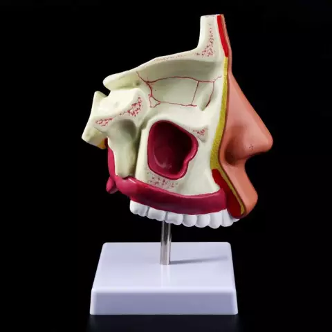
We use every part of our mouth every day, and some of them we even contemplate repeatedly. In science, the structure of the oral cavity is somewhat specified. The photo clearly shows what it is.
Medics in this organ distinguish two sections, called the vestibule of the mouth and its own cavity.
In the vestibule there are external organs (cheeks, lips) and internal (gums, teeth). So to speak, the entrance to the oral cavity is called the oral fissure.
The oral cavity itself is a kind of space, bounded on all sides by organs and their parts. From below - this is the bottom of our oral cavity, from above the palate, in front - gums, as well as teeth, behind the tonsils, which are the border between the mouth and throat, from the sides of the cheek, in the center of the tongue. All internal parts of the oral cavity are covered with mucous membranes.
Lips
This organ, which the weaker sex pays so much attention to rule the stronger sex, is, in fact, a pair of muscle folds surrounding the oral fissure. Atof a person, they are involved in the retention of food entering the mouth, in sound production, in facial movements. The upper and lower lips are distinguished, the structure of which is approximately the same and includes three parts:
- External - covered with keratinizing squamous stratified epithelium.
- Intermediate - has several layers, the outer of which is also horny. It is very thin and transparent. Capillaries perfectly shine through it, which causes the pink-red color of the lips. Where the stratum corneum passes into the mucous membrane, a lot of nerve endings are concentrated (several tens of times more than in the fingertips), so the human lips are unusually sensitive.
- Mucous, occupying the back of the lips. It has many ducts of the salivary glands (labial). Covers it with non-keratinized epithelium.

The mucosa of the lips passes into the mucosa of the gums with the formation of two longitudinal folds, called the frenulum of the upper lip and lower.
The border of the lower lip and chin is the horizontal chin-labial sulcus.
The border of the upper lip and cheeks are the nasolabial folds.
The lips are joined together at the corners of the mouth by labial adhesions.
Cheeks
The structure of the oral cavity includes a paired organ, known to everyone as the cheeks. They are divided into right and left, each has an outer and inner part. The outer is covered with thin delicate skin, the inner is non-keratinizing mucosa, passing into the mucous membrane of the gums. There is also a fatty body in the cheeks. In infants, it performsan important role in the process of sucking, therefore it is developed significantly. In adults, the fat body flattens and moves back. In medicine, it is called Bish's fat lump. The basis of the cheeks are the cheek muscles. There are few glands in the submucosal layer of the cheeks. Their ducts open in the mucous membrane.
Sky
This part of the mouth is essentially a partition between the oral cavity and the nasal cavity, as well as between the nasal part of the pharynx. The functions of the palate are mainly only the formation of sounds. It participates insignificantly in chewing food, as it has lost its clear expression of transverse folds (in infants they are more noticeable). In addition, the palate is included in the articulatory apparatus, which provides bite. Distinguish between hard and soft palate.

2/3 is hard. It is formed by the plates of the palatine bones and processes of the maxillary bones, fused together. If, for some reason, fusion does not occur, the baby is born with an anomaly called a cleft palate. In this case, the nasal and oral cavities are not separated. Without specialized help, such a child dies.
The mucosa during normal development should grow together with the upper palate and smoothly pass to the soft palate, and then to the alveolar processes in the upper jaw, forming the upper gums.
The soft palate accounts for only 1/3 of the part, but it has a significant impact on the structure of the oral cavity and pharynx. In fact, the soft palate is a specific fold of mucous, like a curtain hanging over the root of the tongue. She separates her mouth fromthroats. In the center of this "curtain" there is a small process called a tongue. It helps to form sounds.
From the edges of the "curtain" depart the anterior arch (palato-lingual) and the back (palatopharyngeal). Between them there is a fossa where an accumulation of cells of lymphoid tissue (palatine tonsil) is formed. The carotid artery is located 1 cm from it.
Language
This organ performs many functions:
- chewing (sucking in babies);
- sound-forming;
- salivary;
- taster.

The shape of a person's tongue is influenced not by the structure of the oral cavity, but by its functional state. In the tongue, a root and a body with a back (the side facing the palate) are distinguished. The body of the tongue is crossed by a longitudinal groove, and at the junction with the root lies a transverse groove. Under the tongue is a special fold called the frenulum. Near it are the ducts of the salivary glands.
The mucosa of the tongue is covered with a multi-layered epithelium, which contains taste buds, glands and lymph formations. The top, tip and lateral parts of the tongue are covered with dozens of papillae, which are divided in shape into mushroom-shaped, filiform, conical, leaf-shaped, grooved. There are no papillae at the root of the tongue, but there are clusters of lymphatic cells that form the tongue tonsils.
Teeth and gums
These two interrelated parts have a great influence on the structure of the oral cavity. Human teeth begin to develop during the embryonic stage. Ata newborn in each jaw has 18 follicles (10 milk teeth and 8 molars). They are located in two rows: labial and lingual. The appearance of milk teeth is considered normal when the baby is 6 to 12 months old. The age at which milk teeth normally fall out is even more extended - from 6 to 12 years. Adults should have from 28 to 32 teeth. A smaller number negatively affects the processing of food and, as a result, the work of the digestive tract, since it is the teeth that play the main role in chewing food. In addition, they are involved in the correct sound formation. The structure of any of the teeth (indigenous or milk) is the same and includes the root, crown and neck. The root is located in the dental alveolus, at the end it has a tiny hole through which veins, arteries and nerves pass into the tooth. A person has formed 4 types of teeth, each of which has a certain crown shape:
- cutters (in the form of a chisel with a cutting surface);
- fangs (conical);
- premolars (oval, has a small chewing surface with two tubercles);
- large molars (cubic with 3-5 tubercles).
The necks of the teeth occupy a small area between the crown and the root and are covered by the gums. At their core, gums are mucous membranes. Their structure includes:
- interdental papilla;
- gingival margin;
- alveolar area;
- mobile gum.
The gums consist of stratified epithelium and lamina.
Their basis is a specific stroma, consisting of many collagen fibers that providea snug fit of the mucosa to the teeth and the correct chewing process.

Microflora
The structure of the mouth and oral cavity will not be fully disclosed, if not to mention the billions of microorganisms for which, in the course of evolution, the human mouth has become not just a home, but the whole universe. Our oral cavity is attractive to the smallest bioforms due to the following features:
- stable, moreover, optimal temperature;
- constantly high humidity;
- slightly alkaline medium;
- almost constant availability of freely available nutrients.
Babies are born into the world already with microbes in their mouths, which move there from the birth canal of women in labor in the shortest time until the newborns pass them. In the future, colonization moves at an amazing speed, and after a month of microbes in the mouth of a child, there are several dozen species and millions of individuals. In adults, the number of microbe species in the mouth ranges from 160 to 500, with numbers reaching into the billions. Not the last role in such a large-scale settlement is played by the structure of the oral cavity. Teeth alone (especially diseased and uncleaned ones) and the almost constantly present plaque on them contain millions of microorganisms.
Bacteria prevail among them, the leader among which are streptococci (up to 60%).
Besides them, fungi (mainly candida) and viruses live in the mouth.
Structure and function of the oral mucosa
From the penetration of pathogenic microbes into the tissues of the oral cavityprotected by the mucous membrane. This is one of its main functions - the first to take the hit of viruses and bacteria.
It also covers the tissues of the mouth from exposure to adverse temperatures, harmful substances and mechanical injuries.
In addition to the protective, the mucosa performs another very important function - secretory.
The structural features of the oral mucosa are such that glandular cells are located in its submucosal layer. Their accumulations form small salivary glands. They continuously and regularly moisturize the mucosa, ensuring its protective functions.

Depending on which departments the mucous membrane covers, it can be with a keratinized surface layer or epithelium (25%), non-keratinized (60%) and mixed (15%).
Only the hard palate and gums are covered with keratinized epithelium, because they take part in chewing and interact with solid food fragments.
Non-keratinizing epithelium covers the cheeks, soft palate, its process - the uvula, that is, those parts of the mouth that need flexibility.
The structure of both epithelium includes 4 layers. The first two of them, basal and spinous, both have.
In the keratinized layer, the third position is occupied by the granular layer, and the fourth by the stratum corneum (there are cells without nuclei and practically no leukocytes).
In the non-keratinizing third layer is intermediate, and the fourth is superficial. There is an accumulation of leukocyte cells in it, which also affects the protective functions of the mucosa.
Mixed epithelium covers the tongue.
The structure of the oral mucosa has other features:
- The absence of a muscular plate in it.
- The absence of a submucosal base in certain parts of the oral cavity, that is, the mucosa lies directly on the muscles (observed, for example, on the tongue), or directly on the bone (for example, on the hard palate) and is firmly fused with the underlying tissues.
- The presence of multiple capillaries (this gives the mucosa a characteristic reddish color).
The structure of the oral cavity in children
During a person's life, the structure of his organs changes. So, the structure of the oral cavity of children under one year is significantly different from its structure in adults, and not only by the absence of teeth, as mentioned above.
The primary mouth of the embryo is formed in the second week after conception. Newborns, as everyone knows, do not have teeth. But this is not at all the same as the absence of teeth in the elderly. The fact is that in the oral cavity of babies, teeth are in a state of rudiments, and, at the same time, both milk and permanent teeth. At some point, they will appear on the surface of the gums. In the oral cavity of the elderly, the alveolar processes themselves are already atrophied, that is, there are no teeth and never will be.

All parts of the mouth of a newborn are created by nature in such a way as to ensure the sucking process. Features that help nipple latching:
- Soft lips with specific lip pad.
- Relatively well developed circular muscle inmouth.
- Gingival membrane with many tubercles.
- The transverse folds in the hard palate are clearly defined.
- The position of the lower jaw is distal (the baby pushes his lower jaw, and makes it move back and forth, and not to the sides or in a circle, as when chewing).
An important feature of babies is that they can swallow and breathe at the same time.
The structure of the oral mucosa of infants is also different from that of adults. The epithelium in children up to a year consists of only the basal and spinous layers, and the epithelial papillae are very poorly developed. In the connective layer of the mucosa, there are protein structures transferred from the mother along with immunity. Growing up, the baby loses its immune properties. This also applies to the tissues of the oral mucosa. In the future, the epithelium thickens in it, the amount of glycogen on the hard palate and gums decreases.
By the age of three in children, the oral mucosa has more distinct regional differences, the epithelium acquires the ability to keratinize. But in the connecting layer of the mucosa and near the blood vessels there are still many cellular elements. This contributes to increased permeability and, as a result, the occurrence of herpetic stomatitis.
By the age of 14, the structure of the oral mucosa in adolescents is not much different from adults, but against the background of hormonal changes in the body, they may experience mucosal diseases: mild leukopenia and youthful gingivitis.


