- Author Curtis Blomfield blomfield@medicinehelpful.com.
- Public 2023-12-16 20:44.
- Last modified 2025-01-23 17:01.
The world around us is painted in a variety of colors and shades. Human eyes are able to catch this color variety. For many, it is important to choose clothes in matching colors. For others, it is important to arrange their own interior in pleasant colors. Still others do not understand their life without admiring the beauty and scenic beauty of nature. What would life be like if a person saw everything in black and white? How do people with color blindness see?

Color sensation
The human eye is able to see colors due to the variety of radiation ranges of the light spectrum. The cone apparatus of the retina is responsible for this function.
There are three groups of color waves:
- Long wave - orange and red.
- Mid-wave - green and yellow.
- Shortwave - cyan, violet and blue.
The main colors are red, green and blue. By mixing these colors in various proportions, you can get many shades thatperceives the eye.
Sometimes there are disturbances in the work of cones, and the eye cannot distinguish colors. The male half of the population most often suffers from such disorders.
To determine the pathology of color perception in humans, tables are used to check color perception.
For the first time, the study of the phenomenon of color blindness was started in 1794 by a scientist from England named John D alton. This scientist did not distinguish between the color red, like his two siblings. This vision disorder was named after him.
Colorblindness
The inability of the eyes to distinguish color shades is defined as color blindness.
Scientists have found that there is a congenital disorder of color perception and received in connection with some factors. Men with this pathology are born 16 times more than women.
Color blindness differs in three ways:
- When the inability to clearly distinguish the color red, this condition is called protanomaly (protos - from the Greek. first).
- If the eye's perception of green color is impaired, this is called deuteranomaly (deuteros, Greek for second).
- When the perception of blue color is disturbed, this is tritanomaly (tritos from the Greek third).
In turn, the color blindness of red and green colors is divided into types:
- C - slight deviation from the norm of color perception.
- B - a significant deviation from the norm of color perception.
- A - complete loss of the ability to perceive green or red.
This pathology is determined by the check tablevision and color perception.
Types of color blindness
When you lose the ability to distinguish one of the colors, a person is called a dichromate. A person with normal color perception is called a trichromat.
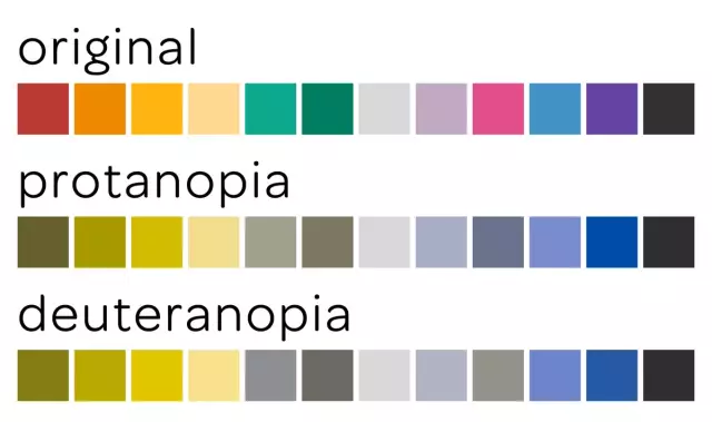
In the complete absence of the perception of red, the pathology is called protanopia, green - deuteranopia, blue - tritanopia. If one of the three colors is not perceived, the perception of the other two is disturbed.
A rare type of color blindness, when a person distinguishes only one color out of three (monochromatic). And the rarest case, in the complete absence of color perception (achromasia), when a person sees everything in black and white.
Visual color discrimination tests use polychromatic tables to test color perception.
Cause of color blindness
Color blindness is not a disease, it is a genetic anomaly that is inherited. The altered gene passes through the female line, but women themselves almost never suffer from color blindness, but their children, boys, are very likely to get this disease.
Color blindness may not occur from birth, but as a result of trauma, surgery or as a reaction to the use of drugs.
All colorblind people see colors differently, depending on the degree of mutation in the cone apparatus of the eyes.
Until the end, the cause of color blindness has not been studied, but it is believed that this is the result of evolution as an adaptation to the environment.
How colorblind people see
It is clear that colorblind people perceive the world differently than people with normal color perception. But,accustomed from birth to just such a vision, they learn to live with it.

Many colorblind people can see colors against a background of another color, while ordinary people see only one color.
On the tables for determining color perception, a color-blind person cannot distinguish the background color of the depicted figure or figure on it by a tone lower or higher. It sees all parts of the image in the same color.
When is color blindness a problem?
A person suffering from a color vision disorder may not be aware of his illness. But there are a number of activities in which it is important that the human eye perceives all three primary colors of the spectrum.
Drivers must distinguish between the color of road signs, parking lights and brake lights on cars of other road users, as well as the colors of traffic lights. That is why when passing a medical examination for obtaining a driver's license, it is mandatory to pass a test using color perception tables for drivers.
Workers in production using special equipment must distinguish color signals.
In medicine, it is very important to distinguish shades and colors for diagnosis and surgery.
It is equally important for a pastry chef to distinguish between shades and colors to create delicious and colorful cakes and pastries.
Methods for diagnosing color blindness
D altonism is usually diagnosed as a result of a routine or random medical examination by an ophthalmologist. The patient is asked to look at the charts to checkcolor perception of Rabkin and Yustova or examine his eyes using the Rabkin Spectral Anomaloscope.

With the help of these studies, it is possible to determine whether this disorder is congenital or acquired.
Tables are square or round pictures, which depict small colored circles in the form of a number or figure against a background of small circles of a different color. Color-blind people see all the circles in the picture of the same color and cannot distinguish the figure or number depicted on it.

Tables for checking color perception
Professor and ophthalmologist Rabkin E. B. in 1936 created his first polychromatic tables for the study of color vision.
These tables allow you to determine the type of color blindness and its complexity. All over the world, these tables are used by ophthalmologists.
Circles of the same brightness form an image, where against the background of some circles others are encrypted in the form of a figure or number.
There are a total of 27 tables defining each individual color vision disorder.

Some hidden figures and numbers are visible to people with good color perception, in other pictures hidden images are visible only to color blind people.
When diagnosing color blindness, tables are often used to check the color perception of Yustova E. N.
Her tables are square pictures, each of which consists of two colors. In the center of one such picture is a square without one wall. The central square and the background are different in color. These pictures are shown as small squares, closely spaced.
To determine Yustova's visual anomaly, 12 variants of pictures were created.
When examining, it is necessary to determine on which side the central square does not have a wall (top, bottom, left, right).
Evaluation of color perception using tables
When examining color perception using Rabkin's technology, polychromatic cards are placed in front of the subject in a well-lit room. The light should fall directly on the pictures. At a distance of half a meter to a meter, the subject must distinguish between the drawings hidden in the plates. One image should take no more than five seconds.
If a visual anomaly test is performed on a child, he is asked to circle the number or shape he sees with a finger or a brush.

If the final conclusion is difficult or there is a suspicion that the subject has memorized the tables with answers to check color perception, there are control tables in Rabkin's set. There are 22 of them. Trichomats with normal vision correctly name all the colors, shapes and numbers indicated on them. Dichromats can only name 10 of them.
To reduce the time spent on this study, it is enough to take three cards with the most difficult image and show them to the subject several times.
In difficult cases, they resort in addition to tables to determinethreshold color vision. With their help, they determine the line when a person stops seeing the hue and saturation of color. It's called color power.
The test is carried out in sufficient light. The subject is asked to look at the tables through a special mask with a round hole. The 12 tables consist of red, yellow, green, blue and grey. On 11 of them there is a scale with options for a smooth transition from white to a rich color tone. On one remaining black and white field, so that the subject knows what to look for.

Tables are counted in order from left to right, top to bottom.
Each card consists of 36 cells arranged in a 6 x 6 square. 26 of them have the main color, and 10 cells, arranged in the form of a "P" or a square without one side, have the same color, but differ in tone. The subject must determine on which side the square does not have a wall. On each subsequent card, the difference between the main color and the central square becomes more noticeable.
The positive side of this study is that it cannot be faked. The subject will not be able to memorize the answers to the cards. Whereas with Rabkin, when examining drivers, the tables for checking color perception with answers will not be difficult to memorize and falsify the results.
The disadvantage of Yustova's tables is the quality of the image and color reproduction, which can be impaired when using low-quality paper or ink from a printing device.
The subject separates each field of vision from the others using a round hole. Fields must be reviewed at least three times each for a valid result.
Results
If in the study of color vision using Rabkin's tables all 27 tables are named correctly, the color vision of the subject is considered correct.
If there is no red color in the spectrum, usually 7 tables are correctly named, in the absence of green - 9 tables, and if the blue color is indistinguishable, 23 tables are correctly named.
When using Yustova's tables, the degree of vision of shades of the same color is determined, which change from more saturated to barely distinguishable. If the perception of red color is impaired, the subject cannot determine the direction "P" on plates 1-4. In violation of green vision, 5-8 tables are not distinguishable. Problems in blue help identify 9-11 tables.
Each table belonging to a certain color group, in order, has its own difference threshold 5 - difficult to distinguish, 10 - less difficult, 15-20 - medium difficulty, 30 - the easiest difference.
Gradual increase in the difficulty of distinguishing between the cells of the tables allows you to identify congenital and incipient deterioration due to color vision disease. They also allow you to control the dynamics of healing.


