- Author Curtis Blomfield blomfield@medicinehelpful.com.
- Public 2023-12-16 20:44.
- Last modified 2025-01-23 17:01.
In this article we will tell you what it is - TKDG, and how the study is conducted.
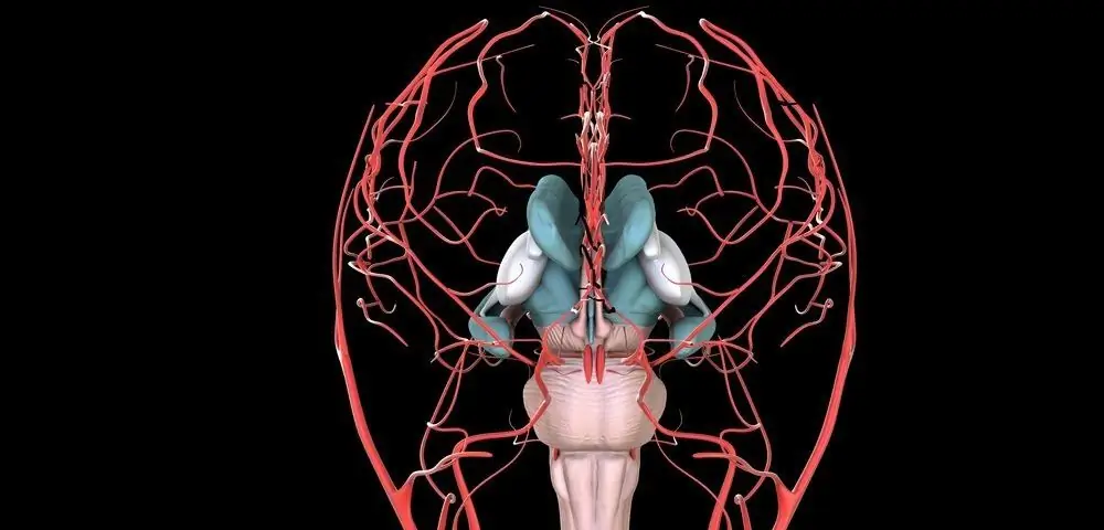
Oxygen and other important elements for life through the arteries and veins enter the cells of the brain and other organs. Nutrient deficiency negatively affects the state of the human body. Ultrasound dopplerography (dopplerography of the cervical vessels, ultrasound, doppleroscopy of cerebral vessels, dopplerography of cerebral vessels, transcranial dopplerography) is a non-invasive method for analyzing blood flow. This procedure is advised by a neurologist in order to prescribe the necessary treatment or undergo additional studies.

The essence of dopplerography
This technique is highly informative, with its help many brain disorders are diagnosed. Doppler ultrasound (USDG) or TKDGhead and neck vessels combines studies based on the Doppler effect and ultrasound diagnostics. From a technical point of view, Dopplerography can be described in a simplified way as follows: reflected high-frequency sound waves affecting blood vessels contribute to the visualization of the structure of veins and arteries, while dopplerography of the head vessels reflects the movement of red blood cells at a particular moment. With the help of a computer, two ready-made images allow you to get an accurate picture of the movement of blood. Due to the color coding, the device provides a full visibility of blood flow and features of existing diseases.
Exploration Modes
The study uses several modes, depending on the specific area:
- scanning of the main arteries, which allows you to study vascular blood flow;
- duplex arterial and venous scanning, which gives the specialist a color two-dimensional sketch, which fixes the state of the vessels in the neck, skull, both lobes of the brain;
- triplex scanning, which complements the first two studies.

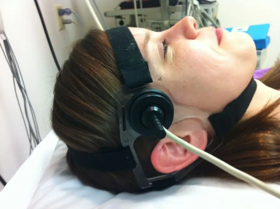
To display comprehensively the state of the vessels of the brain, the main veins and arteries that provide blood flow, these modes are applied simultaneously. Thanks to TKDG of the vessels of the head and neck, a clinical picture is reliably obtained on a computer monitor using the radiation of an ultrasonic wave that penetrates the vessels, is reflected from red blood cells, depending on the speed and direction, inwhich they are moving. Reflected waves are caught by special sensors and converted into an electrical signal, which forms a sketch in dynamics. If there are blood clots, narrowing of the gaps and spasm, the course of the blood changes. This immediately gets a commit on the screen. To study the veins and arteries located in the brain, a transducer is placed on the bones of the skull, which have a minimum thickness.
Triplex Study
Patients often ask the question: how is a triplex study of cerebral vessels carried out, what is its essence and purpose? With ordinary dopplerography, the doctor evaluates only the patency of the vessels, with duplex scanning, the vascular structure, speed and intensity of blood flow are revealed. Triplex TKDG of the vessels of the head and neck allows you to examine the structure of blood vessels, as well as the dynamics of blood movement, to obtain a large-scale picture of vascular patency in color.

Who needs a test?
To understand that there is insufficient nutrition of cells in the brain, you need to pay attention to the following symptoms:
- balance problems, unsteady gait, problems while walking;
- decrease in hearing and vision acuity;
- violation of speech articulation;
- frequent headaches;
- tinnitus;
- vomiting and bouts of nausea not caused by indigestion;
- dizziness, especially when bending over;
- fainting;
- change in taste sensations;
- insomnia;
- memory loss, impaired concentration;
- chillness, swelling and numbness of hands and feet;
- desensitization of certain areas of the body.
Dopplerography of the brain of the head is indicated for patients with certain chronic diseases. The specialist may schedule an examination for people who:
- suffer from diabetes and vascular dystonia;
- smoke;
- are overweight;
- had a heart attack or stroke;
- have cervical osteochondrosis;
- feel very tired when walking, running and light physical activity;
- have palpitations if there is a chance that a broken blood clot will clog an artery that feeds the brain;
- have a pulsating focus in the neck.
In addition, a study can be ordered before heart surgery, as well as for children with mental developmental disorders.
Based on the patient's complaints, the neurologist can determine where the blood flow in the body has been disturbed.

What is determined?
TCDG of head and neck vessels helps:
- determine the narrowing of the arterial lumen and the severity of the diagnosed disorder;
- detect the strength and speed of blood flow in the main arteries;
- detect cerebral aneurysms;
- to assess the condition of the spinal arteries;
- analyze what state the vessels are in when squeezed, bent anddeformations;
- identify disorders that result from the accumulation of atherosclerotic plaques or blood clots;
- find the causes of headaches, which can be circulatory disorders, high intracranial pressure and angiospasms.
Patient preparation
The patient should refrain from drinking coffee, tea, alcoholic and energy drinks the day before the ultrasound and TKDG of the vessels of the head and neck. It is undesirable to smoke at least four hours before the procedure and take medicines. The meaning of such restrictions is that there is no unnecessary load on the vessels and, in general, on blood pressure. When a child undergoes an examination, it is required to ensure his emotional and physical rest.

What is important to understand?
The patient must understand that the study is completely painless, he needs to follow all the doctor's requirements, not to make unnecessary movements and not to talk. The specialist may ask a small patient to hold his breath or start breathing quickly. It is advisable to feed children under one year of age an hour before the study and ensure sleep. There are no restrictions on eating on the eve of the procedure, but you should not eat immediately before it, because when food is digested, blood flow to the head decreases, and the study may be distorted. Please bring a towel for hygiene purposes.
Features
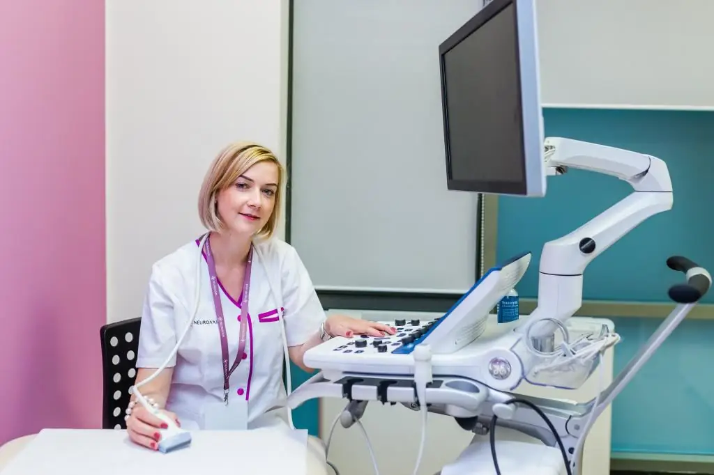
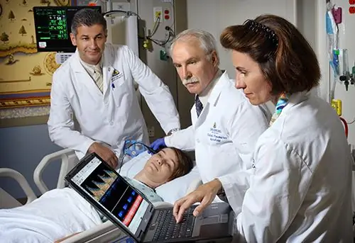
Occupying a horizontal position on the couch,the patient lies on his back. Dopplerography of the vessels of the head and neck with Doppler lasts from 20 to 35 minutes. The specialist applies a special gel on the body to facilitate the sliding of the sensor and avoid interference. First, the neck is analyzed by smoothly moving the sensor along the neck up to the lower jaw. They put it on the points where medium and large veins and arteries are located. The patient is asked several times to hold his breath, and also to change the position of the body in order to accurately assess the vascular tone. After that, the device is switched to the color mode to detect vascular pathologies, areas with impaired blood flow and blood clots.

The study is carried out on the back of the head, temples and scalp. During it, loads on the vestibular apparatus, light flashes, sound stimuli, head turns, breath holding and blinking are used. Vascular dopplerography of the vessels of the brain of the head does not bring pain. When the procedure ends, it will be necessary to carry out hygienic manipulations - remove the remaining gel.
How is the data decrypted?
Deciphering with ultrasound and Dopplerography of the vessels of the head and neck consists in a comparative analysis of the artery with specific norms for a number of parameters: diameter; wall thickness; resistance indices; features of blood flow symmetry; systolic peak velocity. Veins are evaluated according to the vascular diameter, the nature of blood exchange, and the condition of the walls of the veins. If the results are normal, then:
- between the vessels will be clearly visible andfree clearance;
- thickness of he althy arterial walls should be about one centimeter;
- in areas where there is no vascular branching, there is no turbulent flow;
- should also not have arteriovenous malformations;
- when the vessels are squeezed together, an additional examination will be required;
- if any area is abnormal, the doctor indicates an alphanumeric abbreviation;
- knowing all the designations, the neurologist can easily decipher the information received and prescribe the appropriate therapy.
Contraindications and perceived risks
There are no unconditional contraindications that prohibit the procedure. Limitations are the serious condition of the patient and other factors that do not allow the patient to take a supine position (chronic heart failure, exacerbation of bronchial asthma). Dopplerography is absolutely safe for the human body, it does not carry radiation exposure. This procedure has no age restrictions. It is carried out for the elderly, infants, nursing mothers, pregnant women. During pregnancy, the doctor may refer for research if there is a possibility of circulatory disorders in the placenta.

TCDG DS of the vessels of the head and neck is allowed to be carried out repeatedly. Due to the harmlessness and high accuracy, the technique is used to control the ongoing treatment. In addition, it is also prescribed for the prevention of serious diseases.
During the study, certaindifficulties: it is more difficult to assess the condition of small vessels than large ones; it happens that the cranial bones interfere with the full analysis of the state of all the vessels of the brain. The results often depend on the skill of the specialist and the instruments. Often transcranial doppler sonography (TCD) is used instead of angiography.


